Pulpectomy is an essential dental procedure aimed at preserving primary teeth affected by severe infections or irreversible pulpitis. Zinc oxide eugenol (ZOE), a commonly used obturating material, presents limitations including slow resorption, cytotoxicity, and potential adverse effects on developing permanent teeth. In response to these challenges, a novel obturating material combining zinc oxide, calcium hydroxide, and metronidazole has been developed. This study aimed to compare the cytotoxicity of the novel obturating material with that of ZOE using an MTT assay. Cytotoxicity was assessed using mouse fibroblast cells (3T3-L1) cultured in DMEM-F12 medium. The cells were treated with various concentrations of the novel material and ZOE, followed by MTT assay to measure cell viability. Absorbance was recorded at 570 nm, and statistical analysis was conducted using SPSS software. At lower concentrations (2.5 to 5 µl/ml), both materials exhibited minimal cytotoxicity, with cell viability exceeding 90%. However, as the concentration increased, both materials demonstrated a dose-dependent decline in cell viability. At 60 µl/ml, the novel material exhibited 56.63% cell viability, whereas ZOE showed 45.36%, indicating greater cytotoxicity associated with ZOE at higher concentrations. Intra-group analysis revealed significant cytotoxic differences between low and high concentrations for both materials. The novel obturating material demonstrates lower cytotoxicity than ZOE, particularly at lower concentrations, positioning it as a safer and more biocompatible alternative for pulpectomy in pediatric dentistry. Further clinical trials are warranted to validate these findings and evaluate long-term outcomes.
Introduction
Pulpectomy is a dental procedure designed to preserve primary teeth that are significantly infected or exhibit irreversible pulpitis [1]. It procedure involves the complete removal of necrotic or inflamed pulp tissue from the root canals, followed by cleaning, shaping, and obturation (filling) of the root canal space [2]. The primary objective of pulpectomy is to eliminate the infection, prevent reinfection, and maintain the tooth's function until it naturally exfoliates and is replaced by the permanent tooth [3-6]. This procedure is particularly crucial in pediatric dentistry, as the early loss of primary teeth can lead to developmental issues, such as malocclusion and speech difficulties [7, 8].
The success of root canal therapy in primary teeth largely depends on the selection of obturating material [9-14]. An ideal root canal filling material for primary teeth should possess strong antibacterial properties and resorb at a rate commensurate with that of the primary teeth, without adversely affecting the permanent teeth or surrounding periapical tissues [15-27]. Zinc oxide eugenol (ZOE) was the first obturating material introduced for primary teeth, recognized for its anti-inflammatory and analgesic properties, and has been regarded as the gold standard for obturation in primary teeth [28, 29]. However, ZOE can irritate periapical tissues and tends to resorb slowly, potentially interfering with the eruption of permanent teeth [30]. Additionally, once set, ZOE becomes brittle, which may result in poor adaptation to the irregularities of the root canal walls, particularly in curved or accessory canals, creating voids that increase the risk of reinfection [31]. Furthermore, when extruded beyond the root apex, ZOE can harm the developing permanent tooth, leading to developmental defects [32]. Eugenol, a component of ZOE, can also trigger allergic reactions in some patients, resulting in localized irritation, discomfort, or hypersensitivity reactions [33-35]. This limitation restricts its use in certain populations, especially in children with unknown sensitivities to eugenol [36].
To address these limitations while leveraging the benefits of zinc oxide eugenol, a novel obturating material was developed, combining zinc oxide, calcium hydroxide, and metronidazole. The slower resorption rate of zinc oxide [37-39].
Materials and Methods
Study design and ethical approval
This research was designed as an in vitro study to compare the cytotoxicity of a newly developed obturating material with Zinc Oxide Eugenol, utilizing the MTT Assay. Ethical approval was obtained from the Institutional Review Board of Saveetha Dental College and Hospitals prior to the commencement of the study (IHEC/SDC/PEDO-2205/24/071).
Preparation of the obturating material
A 60-40 concentration was achieved by combining 600 mg of calcium hydroxide with 400 mg of powdered zinc oxide. The mixture was stirred for 60 minutes using a magnetic stirrer to ensure uniformity. Metronidazole tablets (400 mg) were ground into a powder using a mortar and pestle. The final obturating material, with a 2% concentration, was prepared by mixing 4 mg of metronidazole powder with 196 mg of the calcium hydroxide-zinc oxide mixture.
Cell culture
Mouse fibroblast cells (3T3-L1) were cultured in Dulbecco's Modified Eagle Medium F-12 (DMEMF-12) supplemented with a 1X antibiotic solution (streptomycin and penicillin) and 10% fetal bovine serum (FBS). The cells were incubated at 37°C with 5% CO₂ for optimal growth conditions for 24 hours. Following incubation, the cells were washed with 100 μL of 1X Phosphate Buffered Saline (PBS) and treated with buffalo fat, followed by an additional 24-hour incubation under the same conditions.
MTT cell viability assay
At the conclusion of the treatment period, the medium was aspirated, and 0.5 mg/mL of 3-(4,5-dimethylthiazol-2-yl)-2,5-diphenyltetrazolium bromide (MTT) solution prepared in 1X PBS was added to each well. The cells were incubated for 4 hours at 37°C. After incubation, the MTT solution was discarded, and the cells were rinsed with 100 μL of PBS. The formazan crystals formed were dissolved in 100 μL of Dimethyl Sulfoxide (DMSO), and absorbance was measured at 570 nm using a microplate reader. The percentage of cell viability was calculated using the following formula:
|
Cell viability (%)=(O.D. of control cells)(O.D. of treated cells)×100 |
(1) |
Morphological analysis
The IC₅₀ value for both compounds, as determined from the MTT assay results, was established at 20 μM/mL, which was subsequently utilized for further morphological analysis. Mouse fibroblast cells (2 × 10⁵) were cultured in 6-well plates and treated with 20 μM/mL of the selected compounds for a duration of 24 hours. Following incubation, the medium was aspirated, the cells were rinsed with PBS (pH 7.4), and cell morphology was assessed using a phase-contrast microscope.
Statistical analysis
Data entry and statistical analysis were conducted using SPSS software (IBM SPSS Statistics, Version 21.0, IBM Corp., Armonk, NY). The viability of the cells at various concentrations of Zinc Oxide Eugenol and the novel obturating material was compared using an independent t-test. For intra-group analysis, post Hoc Bonferroni and repeated measures ANOVA were utilized, with a p-value greater than 0.05 considered statistically significant.
Results and Discussion
No statistically significant difference was observed in the viability of the cells between the novel obturating material and Zinc Oxide Eugenol across the various concentrations, as illustrated in Table 1.
Table 1. Comparison of cell viability between different concentrations of the novel obturating material and Zinc Oxide Eugenol (Independent t-test, p < 0.05 statistically significant).
|
Group |
N |
Mean |
Std. Deviation |
p-value |
|
|
Control |
Novel Obturating material |
3 |
100.0000 |
.00000a |
1.00000 |
|
ZOE |
3 |
100.0000 |
.00000a |
||
|
2.5 |
Novel Obturating material |
3 |
98.3841 |
1.70646 |
.505 |
|
ZOE |
3 |
97.3706 |
1.68303 |
||
|
5 |
Novel Obturating material |
3 |
97.6958 |
1.66885 |
.125 |
|
ZOE |
3 |
94.5331 |
2.28806 |
||
|
10 |
Novel Obturating material |
3 |
93.1532 |
2.70097 |
.694 |
|
ZOE |
3 |
94.0141 |
2.26913 |
||
|
20 |
Novel Obturating material |
3 |
84.8604 |
5.21164 |
.418 |
|
ZOE |
3 |
87.8749 |
2.50701 |
||
|
40 |
Novel Obturating material |
3 |
74.5749 |
3.30333 |
.108 |
|
ZOE |
3 |
66.6241 |
5.78804 |
||
|
60 |
Novel Obturating material |
3 |
56.6373 |
6.57646 |
.093 |
|
ZOE |
3 |
45.3613 |
6.00065 |
||
Intra-group analysis of the novel obturating material at varying concentrations revealed a statistically significant difference exclusively between the 2.5 μl/ml and 5 μl/ml concentrations. No significant differences were observed among the other concentrations (Tables 2 and 3).
Table 2. Comparison of cell viability across different concentrations of the novelobturating material, p < 0.05 - statistically significant
|
Different concentrations of the novel obturating material |
Mean + SD |
p-value |
|
|
2.5 |
98.384+ 0.985 |
0.04 |
|
|
5 |
97.696+ 0.964 |
||
|
10 |
93.153+1.559 |
||
|
20 |
84.860+3.009 |
||
|
40 |
74.575+1.907 |
||
|
60 |
56.637+3.797 |
Table 3. Post Hoc Pairwise Bonferroni comparison of the Novel Obturating Material
|
(I) factor1 |
Mean Difference (I-J) |
Std. Error |
p-value |
|
|
2.5 |
5 |
.688* |
.022 |
.015 |
|
10 |
5.231 |
.574 |
.178 |
|
|
20 |
13.524 |
3.443 |
.887 |
|
|
40 |
23.809 |
1.433 |
.055 |
|
|
60 |
41.747 |
4.020 |
.137 |
|
|
5 |
10 |
4.543 |
.596 |
.252 |
|
20 |
12.835 |
3.433 |
.970 |
|
|
40 |
23.121 |
1.436 |
.058 |
|
|
60 |
41.058 |
4.011 |
.141 |
|
|
10 |
20 |
8.293 |
3.776 |
1.000 |
|
40 |
18.578 |
1.445 |
.090 |
|
|
60 |
36.516 |
4.272 |
.201 |
|
|
20 |
40 |
10.285 |
4.807 |
1.000 |
|
60 |
28.223 |
6.660 |
.771 |
|
|
40 |
60 |
17.938 |
2.918 |
.382 |
In the Zinc Oxide Eugenol group, a statistically significant difference was observed between the following concentrations: 2.5 μl/ml and 60 μl/ml, 5 μl/ml and 10 μl/ml, 5 μl/ml and 20 μl/ml, 10 μl/ml and 20 μl/ml, as well as 40 μl/ml and 60 μl/ml (Tables 4 and 5).
Table 4. Comparison of cell viability across different concentrations of Zinc Oxide Eugenol, p < 0.05 - statistically significant.
|
Zinc oxide eugenol at Different concentrations |
Mean + SD |
p-value |
|
|
2.5 |
97.371+0.972 |
0.04 |
|
|
5 |
94.533 +1.321 |
||
|
10 |
94.014 +1.310 |
||
|
20 |
87.875 +1.447 |
||
|
40 |
66.624 +3.342 |
Table 5. Post Hoc Pairwise Bonferroni comparison of Zinc oxide eugenol
|
(I) factor1 |
Mean Difference (I-J) |
Std. Error |
p-value |
||
|
2.5 |
5 |
2.837 |
.837 |
1.000 |
|
|
10 |
3.357 |
.837 |
.854 |
||
|
20 |
9.496 |
1.053 |
.181 |
||
|
40 |
30.747 |
3.093 |
.150 |
||
|
60 |
52.009 |
3.120 |
.054 |
||
|
5 |
10 |
.519* |
.015 |
.012 |
|
|
20 |
6.658* |
.220 |
.016 |
||
|
40 |
27.909 |
3.921 |
.288 |
||
|
60 |
49.172 |
3.932 |
.095 |
||
|
10 |
20 |
6.139* |
.218 |
.019 |
|
|
40 |
27.390 |
3.923 |
.299 |
||
|
60 |
48.653 |
3.935 |
.097 |
||
|
20 |
40 |
21.251 |
4.140 |
.539 |
|
|
60 |
42.514 |
4.151 |
.141 |
||
|
40 |
60 |
21.263* |
.345 |
.004 |
|
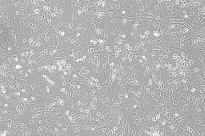
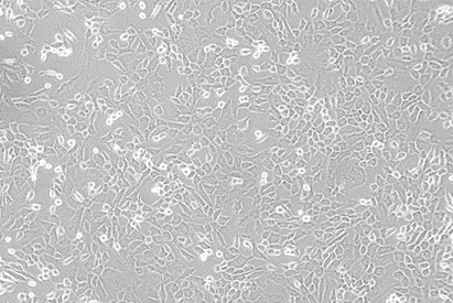
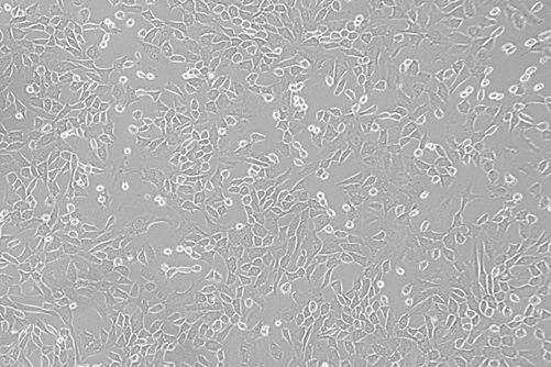
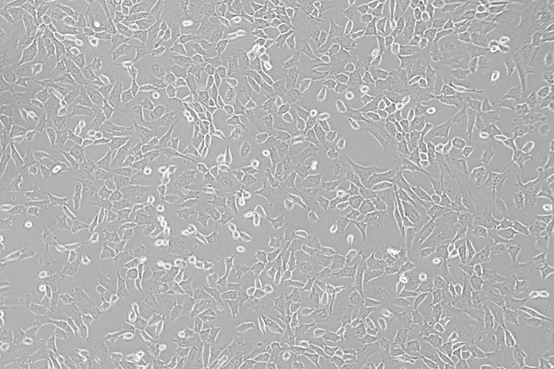
Mouse fibroblast cells were observed under an inverted phase contrast microscope for 24 hours at concentrations of 20 μl/ml and 60 μl/ml. At the lower concentration of 20 μl/ml, both materials exhibited minimal cytotoxic effects (Figure 1), whereas the higher concentration of 60 μl/ml demonstrated increased cytotoxicity (Figure 2).
|
|
|
a) |
|
|
|
b) |
|
Figure 1. Effect of novel obturating material and zinc oxide eugenol on the cell morphology of mouse fibroblasts (3T3-L1). Cells were treated with 20 μl/ml for 24 hours and subsequently observed under an inverted phase contrast microscope. |
|
|
|
a) |
|
|
|
b) |
|
Figure 2. Impact of novel obturating material and zinc oxide eugenol on the cell morphology of mouse fibroblasts (3T3-L1). Cells were treated with a concentration of 60 μl/ml for 24 hours and subsequently observed under an inverted phase contrast microscope. |
Pediatric dentistry plays a crucial role in maintaining deciduous teeth, which are essential for preserving the dental arch and ensuring proper oral function. Ongoing advancements in materials and techniques for pulpectomy are aimed at preserving and protecting the underlying tooth structure [40-43]. A recently developed innovative obturating material is designed to support the preservation of the roots of primary teeth and the underlying permanent tooth buds.
The MTT assay is a widely utilized method in biological research for assessing cell viability, cytotoxicity, and proliferation [44]. In this study, we compare the cytotoxic effects of the novel obturating materials and Metapex using the MTT assay. At lower concentrations (2.5, 5, and 10 µg/mL), both materials exhibited minimal cytotoxicity compared to higher concentrations (20, 40, and 60 µg/mL). This suggests a concentration-dependent reduction in cell viability for both materials, with notable differences emerging as the concentration increases.
Within the novel obturating group, results indicate a significant decrease in cell viability as the concentration of the obturating material increased. At the lowest concentration (2.5 μL/mL), cell viability was highest, with a statistically significant p-value (p = 0.005). However, as the concentration increased to 60 μL/mL, cell viability gradually decreased. This pattern suggests that higher concentrations of the material have a more pronounced cytotoxic effect on mouse fibroblast cells. The findings indicate that the material demonstrates biocompatibility at lower concentrations. A significant mean difference was observed between the 2.5 μL/mL and 5 μL/mL groups (p = 0.015), although the difference was minor, indicating minimal cytotoxicity at these lower concentrations. Comparisons involving higher concentrations (20, 40, and 60 μL/mL) generally exhibited larger mean differences; however, these differences were not statistically significant.
In the zinc oxide eugenol (ZOE) group, cell viability was highest at the lowest concentration of 2.5 μl/ml, with a statistically significant reduction in viability observed as the concentration increased (p = 0.04). At the highest concentration of 60 μl/ml, cell viability decreased markedly. These results indicate that ZOE exhibits greater biocompatibility at lower concentrations, while higher concentrations are associated with significant cytotoxic effects. The pronounced decline in viability at concentrations exceeding 20 μl/ml, particularly at 40 μl/ml and 60 μl/ml, suggests that ZOE's cytotoxic properties become more evident as its concentration increases. This finding is consistent with the known limitations of ZOE, including irritation to surrounding tissues and potential adverse effects on cell health at elevated concentrations.
At lower concentrations, both materials demonstrated cell viability exceeding 90%, indicating their safety and reduced cytotoxicity for clinical use. The novel obturating material exhibited superior cell viability compared to zinc oxide eugenol at lower concentrations, up to 5 µg/mL. However, as the concentration increased, both materials began to exhibit heightened cytotoxic activity. At a concentration of 60 µg/mL, cell viability declined further, with the novel obturating material showing a cell viability of 56.63%, while zinc oxide eugenol demonstrated a cell viability of 45.36%. These findings suggest that zinc oxide eugenol is more cytotoxic than the novel obturating materials at higher concentrations.
A study conducted by Solmaz Mohammadi Nejad et al. raised concerns regarding the cytotoxic effects of eugenol, a component of zinc oxide-eugenol (ZOE) [45]. Eugenol has been demonstrated to induce inflammation and inhibit cell growth, particularly at elevated concentrations, which may be detrimental in clinical settings where tissue regeneration is desired [46]. These findings suggest that while ZOE continues to be a widely utilized material, its cytotoxic effects, especially at higher doses, may limit its long-term clinical safety [47].
Continuous cell lines, such as 3T3 mouse fibroblasts, are routinely employed in the assessment of the cytotoxic effects of various dental materials due to their reproducible growth rates and biological responses [48, 49]. A study by Thonemann et al. indicated that mouse-induced pluripotent stem (iPS) cells exhibit characteristics akin to embryonic stem (ES) cells, including morphology, proliferation, and gene expression in vitro, as well as teratoma formation in vivo [50]. Furthermore, they possess the capacity to produce viable chimeras when injected into blastocysts, confirming germline transmission. Consequently, mouse fibroblasts are utilized to evaluate the cytotoxic effects of obturating materials.
However, the study did not replicate clinical scenarios such as the extrusion of material into periapical tissues, interactions with saliva, or the presence of an active infection—factors that could influence the cytotoxicity of the materials. Additionally, the cytotoxic effects of the materials were not compared with clinical outcomes in patients undergoing pulpectomy, limiting the applicability of the findings to clinical practice. Therefore, further clinical trials are necessary to ascertain whether the novel obturating material offers advantages in terms of ease of use, long-term outcomes, or patient comfort, particularly in pediatric cases.
Conclusion
The novel obturating material exhibits significantly lower cytotoxicity compared to zinc oxide eugenol, particularly at reduced concentrations, thereby presenting a safer and more biocompatible option for clinical use in pulpectomy procedures.
While the performance of the novel obturating material at higher concentrations remains cytotoxic, it may serve as a superior alternative to ZOE due to its combination of antibacterial properties and a more controlled resorption rate. This characteristic could be especially beneficial in pediatric pulpectomy treatments, where excessive resorption or heightened cytotoxicity could adversely affect both deciduous and permanent teeth. Nevertheless, it is essential to continue refining the formulation to further decrease its cytotoxicity at elevated concentrations, as this will enhance its overall safety and efficacy in clinical applications.
Acknowledgments: None
Conflict of interest: None
Financial support: None
Ethics statement: None