2020 Volume 8 Issue 1
Creative Commons License

Prevalence of Congenitally Missing Teeth (Radiographic Study)

Hussein Haleem Jasim

Department of Oral Diagnosis, College of Dentistry, University of Wasit, Kut city, Iraq.


ABSTRACT

The congenitally missing teeth considered a wide distributed among different countries of the world. The people suffered from different problems that are accompanying these anomalies as aesthetic, functional and psychogenic problems. Some factors might be related to the etiology of these cases and variables from hereditary, racial, gender and environmental causes.

Aim of the study: To explore the prevalence of congenitally missing teeth in a sample of the Iraqi population and their correlation to the gender and jaws.

Materials and Methods: The study was undertaken on 395 patients (189 Males and 206 Females) aged ranged between 13 to 25 years who referred to some dentistry centers in Baghdad for taking orthopantomogram (O.P.G) for the period from December 2018 to November 2019. All patients with O.P.Gs were examined clinically on the dental chair for diagnosis of the missing teeth and taking the history to confirm the congenital absence of teeth. The radiographs were viewed by an X-ray viewer. A tooth was registered as a congenitally missing tooth when there was no evidence of any calcification in the missing tooth region on O.P.G radiograph, as well as when the missing tooth region is not due to tooth extraction or exfoliation.

Results: According to the statistical analysis, the rate of congenitally missing teeth in the current study was 10.37% of the study sample. The results showed that most congenitally missing teeth were the maxillary lateral incisors in the percentage of 4.81%, followed by the mandibular second premolars (4.30 %), mandibular lateral incisors (0.50%), maxillary central incisors (0.50%), and maxillary second premolars (0.25 %) respectively. The study showed no type of teeth a genesis for the other teeth. The results showed no significant increase in the prevalence of congenitally missing teeth in females compared with males. The statistical analysis showed no significant increase in the prevalence of congenitally missing teeth in the upper jaws compared with lower jaws. Also, the result of the study showed did not reveal a significant increase in the prevalence of congenitally missing teeth in the left side of the jaws when compared with the right side. The study also showed no significant increase in the prevalence of unilateral congenital missing teeth compared with the bilateral congenitally missing teeth.

Conclusion: The rate of congenitally missing teeth in the sample of the Iraqi population was 10.37%.

Key words: Prevalence, Congenital, Missing, hypodontia, Anadontia, Oligodontia, Teeth.


Introduction


Patients refer to dentists due to different problems.1 There are teeth abnormalities with different types and severities 2-4 such as extra or missing teeth, and Taurodontism.5 The congenitally missing teeth “(teeth agenesis)” are one of the abnormalities that can be occurred in one or both of the jaws which are referred to as an absence of a single tooth or multiple teeth and it may be unilateral or bilateral tooth/ or teeth missing. In general, the congenitally missing teeth interfere negatively on the dental esthetic, occlusion and the masticatory function in the individuals with this abnormality.

Various terms describe the absence of teeth as “hypodontia, oligodontia, and anodontia”.5-8 “Hypodontia” refers to the absence of up to six teeth, “oligodontia” refers to the absence of more than six teeth while “anodontia” refers to the complete absence of teeth.9 That is widely registered in different regions of the world according to many studies.10-29

Studies on this abnormality were distributed around the continents of the world. The studies in Europe, America, and Australia found 5.5% in Europe, 6.3% in Australia and lower rates were in North America.26 These anomalies are rarely associated with deciduous teeth, but their occurrence is common between the permanent teeth, some studies stated that there was a relationship between the congenitally missing teeth in both deciduous and permanent teeth. The reports stated that the occurrence of congenitally missing teeth in children was due to the loss of the corresponding successor's primary teeth.27, 28 Many studies stated that the congenitally missing teeth can be associated with some oral or other abnormalities, “microdontia” considered the most widely feature associated with cases of teeth agenesis.29

Also, other abnormalities may be associated with these cases, like cleft lip and palate, and Down ‘syndrome.6, 27, 30 The ectopic position of permanent, Peg shaped maxillary lateral incisors, enamel hypoplasia, palatal inclined or impacted maxillary canines and molar infra-occlusion.31-33 Rotations and generalized spacing of the teeth beside missing mandibular second premolars can be seen.32 As well as, over-erupted and reclined mandibular incisors which cause increase overbite can be seen.34

Etiology of congenitally missing teeth

During the early phases of tooth development, any disturbances in the early phases of this period lead to congenitally missing teeth.35 Genetics plays a major and critical role in the development of these anomalies.36 Several studies confirmed the genetic effect of researches on monozygotic twins.18, 37-39 It is reported that the absence of anterior teeth may depend mainly on genes, but the absence of posterior teeth might be controversial.40 Environmental factors have a role in the etiology of these anomalies or combination with genetic factors. 

Many studies reported that the “ectoderm” undergoes “dysplastic expression” due to these factors.18, 40-42 Due to the environmental factors, the tooth germ is developing after closing the space available for the development of teeth b the surrounding tissues.20, 43 When the primary teeth are missing congenitally, their permanent analogs may also be missing.18, 44

Various genes were reported and might be responsible for the occurrence of congenitally missing teeth.45-50 Mutation of these genes (as PAX9, TGFA, and MSX) might be associated with the development of congenitally missing teeth between different ethnic groups.15, 49, 51-57 MSX1 and MSX2 have an important role as mediator for the interactions of epithelial and mesenchymal tissues during dental development. 52, 58, 59 For example, mutations in MSX1 have a direct effect on the development of second premolars and third molars and sometimes the first molar.22 But on the common incisor-premolar missing type, MSX1 plays a less important role in the development of this missing type. 46, 58 Moreover, PAX9 and TGFA have a role in the development of congenitally missing teeth by MSX1 and PAX9 interaction.49-52

The understood of the exact genetic mechanism responsible for the development of congenital teeth missing is still vague.20, 42, 51, 60 So independent mechanisms might recommend for each missing tooth.20 Nowadays, mutations in specific genes as LRP6 gene “gene encoding low-density lipoprotein receptor-related protein” has been reported for the etiology of the tooth absence.61

Materials and Methods

The study was undertaken on 395 patients (189 Males and 206 Females) aged ranged between 13 to 25 years who referred to some dentistry centers in Baghdad for taking “orthopantomogram” (O.P.Gs) for the period from December 2018 to November 2019.

All patients with O.P.Gs were examined clinically on the dental chair for diagnosis of the missing teeth and taking the history to confirm the congenital absence of teeth. The radiographs were viewed by an X-ray viewer.

A tooth was registered as a congenitally missing tooth when there was no evidence of any calcification in the missing tooth region on O.P.G radiograph, as well as when the missing tooth region is not due to tooth extraction or exfoliation. Figure 1 & 2.

All patients were informed of the purpose of the study with signed consents.

Exclusion criteria

  • Patients have a dental extraction
  • Patients have previous missing teeth due to trauma or periodontal problems 
  • Patients have cleft lip and palate
  • Patients have down syndrome or any maxillofacial malformations.
  • Third molars were excluded from the study


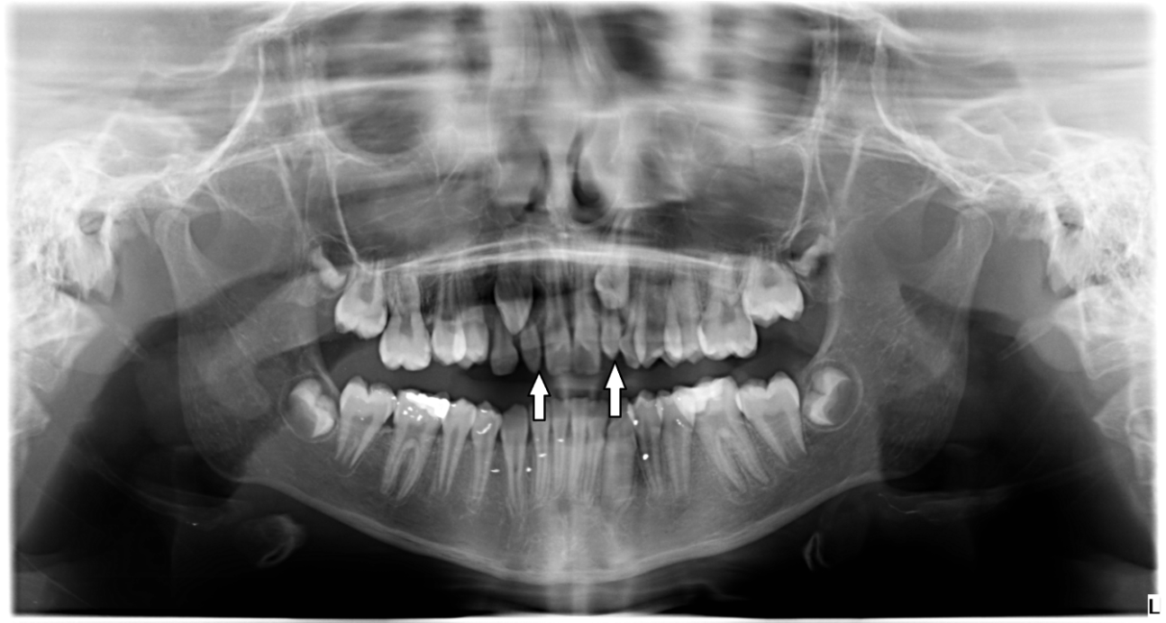
Figure 1. A panoramic radiograph (O.P.G) showed bilateral congenitally missing upper lateral incisors.

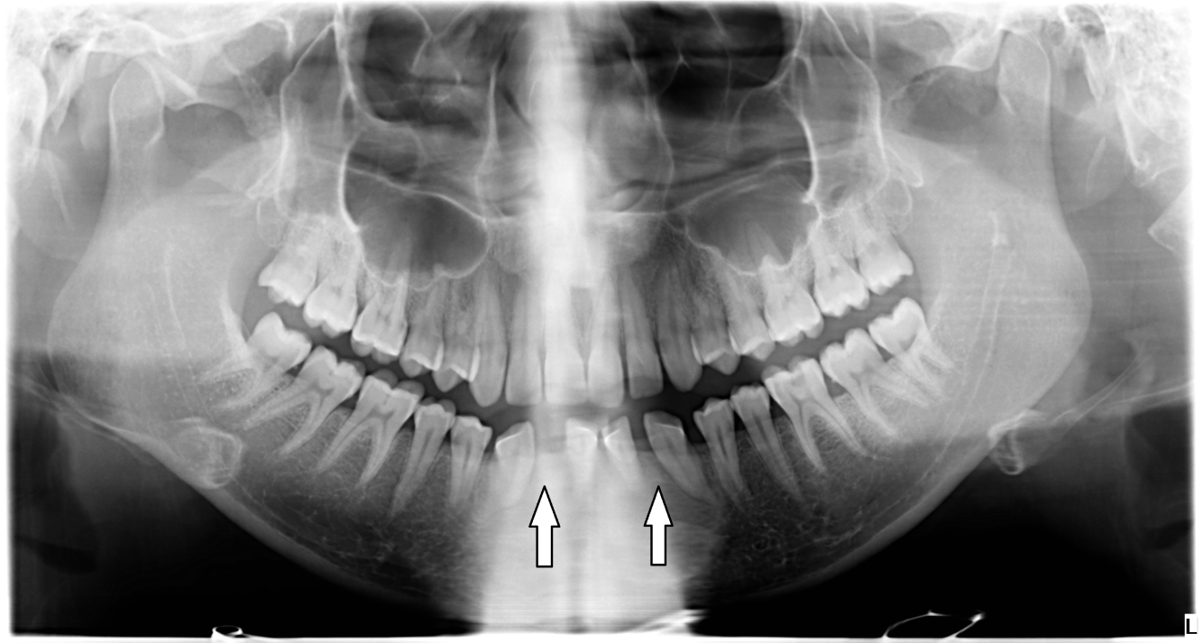
Figure 2. A Panoramic radiograph (O.P.G) showed bilateral congenitally missing lower lateral incisors.


Statistical Analysis

All data were analyzed using the Statistical Package for the Social Sciences software (version 19). The relation between the prevalence of CMT and gender, upper and lower jaws were analyzed using Pearson’s chi-squared test. The significance level was set at 0.05 and considered significant if the P-value  < 0.05.

Results

The statistical analysis showed that most congenitally missing teeth in the current study were the maxillary lateral incisors in the percentage of 4.81%, followed by the mandibular second premolars (4.30%), mandibular lateral incisors (0.50%), maxillary central incisors (0.50%), and maxillary second premolars (0.25%) respectively. [Table 1] The study showed no any type of teeth agenesis for the other teeth. The results showed no significant increase in the prevalence of congenitally missing teeth in females (in the percentage of 11.16%) in comparison with males (9.52%). [Table 2] The results also showed no significant increase in the prevalence of congenitally missing teeth in the upper jaws (in the percentage of 5.56%) in comparison with lower jaws (4.81%). [Table 3] The study also showed no significant increase in the prevalence of unilateral congenital missing teeth (in the percentage of 3.16%) in comparison with the bilateral congenitally missing teeth (2.02%). [Table 4] Also, the study showed no significant increase in the prevalence of congenitally missing teeth on the left side of the jaws (in the percentage of 5.56%) in comparison with the right side (4.81%). [Table 5] According to these results, the rate of congenitally missing teeth in the current study was 10.37% of the study sample.


Table 1. Distribution of CMT in the maxilla and mandible.

Congenitally missing teeth (CMT)

Maxillary arch

Mandibular arch

Males

Females

Total

Males

Females

Total

Right central incisor

0

1

1

0

0

0

Right lateral incisor

4

3

7

0

1

1

Right canine

0

0

0

0

0

0

Right first premolar

0

0

0

0

0

0

Right second premolar

0

1

1

5

4

9

Right first molar

0

0

0

0

0

0

Right second molar

0

0

0

0

0

0

Left central incisor

0

1

1

0

0

0

Left lateral incisor

4

6

12

0

1

1

Left canine

0

0

0

0

0

0

Left first premolar

0

0

0

0

0

0

Left second premolar

0

0

0

3

5

8

Left first molar

0

0

0

0

0

0

Left second molar

0

0

0

0

0

0

Table 2. showed the frequency of CMT in males and females

Gender

Number of CMT

Percentage of CMT

Chi-square test

Significance level at 0.05

Males

18

9.52%

1.24

The p-value is 0.2

Not significant at p-value  < 0.05

Females

23

11.16%


CMT: Congenitally missing teeth


Table 3. showed the frequency of CMT in maxilla and mandible

Type of jaw

Number of CMT

Percentage of CMT

Chi-square test

Significance level at 0.05

Maxillary jaw

22

5.56%

0.23

The p-value is 0.6

Not significant at p-value < 0.05

Mandibular jaw

19

4.81%


CMT: Congenitally missing teeth


Table 4. showed the frequency of unilateral and bilateral CMT in the jaws

CMT in both jaws

Number of CMT

Percentage of CMT

Chi-square test

Significance level at 0.05

Unilateral CMT

25

3.16%

2.08

The p-value is 0.1

Not significant at p-value < 0.05

Bilateral CMT

16

2.02%


CMT: Congenitally missing teeth


Table 5. showed the frequency of CMT in the left and right side of the jaws.

Side of jaw

Number of CMT

Percentage of CMT

Chi-square test

Significance level at 0.05

Left side

22

5.56%

0.23

The p-value is 0.6

Not significant at p-value < 0.05

Right side

19

4.81%


CMT: Congenitally missing teeth


Discussion

A congenitally missing tooth or tooth agenesis is considered to be one of the most anomalies in jaws compared to other dental anomalies that are distributed widely among the people of the world. 

Many authors indicated that several factors could have a role in the incidence of dental anomalies, these factors varied as hereditary, environmental, the pattern of nutrition, chewing function and systemic problems.62-67

Several studies observed the prevalence of congenitally missing teeth all over the world. Graber stated that the ratio of congenitally missing teeth excepting the third molars were 1.6-9.6% in several studies in many countries.41 Other studies stated that the ratio was 2-16.3%.68-70 Also, the ratios in German and Malaysian populations were 12.6% and 2.8% respectively.49, 60 Sheikhi reported that the prevalence of congenitally missing teeth in the Iranian population was 10.9%.57

It was reported that the “teeth agenesis” occurred with the permanent teeth about two to nine times more than the deciduous teeth.18, 58

The prevalence of congenitally missing teeth found with high variation and distribution relative to gender, ethnicity, and race.26, 58, 69, 71-73 For example, Edward and Larkin studied the racial prevalence of congenitally missing teeth in black and white American people and the found that the prevalence was significantly more in white people compared to the black people.74

Other studies reported that environmental and genetic circumstances have an important role in the occurrence of the congenitally missing teeth. 41, 75

The current study observed the prevalence of congenitally missing teeth in a sample of the Iraqi population and also the correlation of the congenitally missing teeth prevalence between genders, upper and lower jaws, and as well as between the right and left side of the same jaw.

The results of current studies showed that most congenitally missing teeth in the current study were the maxillary lateral incisors, followed by the mandibular second premolars, mandibular lateral incisors, maxillary central incisors, and then maxillary second premolars. The study showed no type of “teeth agenesis” for the other teeth.

Many studies have agreed that maxillary lateral incisors are the most congenitally missing teeth followed by the mandibular second premolars.18, 60, 76-79

In many people also it has been reported that the most frequent congenitally missing teeth were the maxillary lateral incisors and then mandibular and maxillary second premolars respectively (except third molars).18, 60, 76, 77

The maxillary lateral incisor also reported being the most common congenitally missing tooth in some studies.20, 36 Also, the maxillary lateral incisor was reported to be the most prevalent congenitally missing tooth in several studies in Iranian12, Turkish,16 Brazilian,15 Sudanese,80 and Malaysian population.60

But on the other hand, some authors reported that the most prevalent congenitally absent teeth were, the mandibular lateral incisors,71 the mandibular central and lateral incisors.26 In another study in the orthodontic Iranian population, the most frequent congenitally missing teeth were the mandibular second and then the maxillary second premolars.57

Several studies also showed that the mandibular second premolars are the most congenitally missing teeth followed by the maxillary lateral incisors and maxillary second premolars.81-84

In some studies also it was reported that the mandibular second premolars were the most frequent congenitally missing teeth and then the maxillary lateral incisors and the mandibular central incisors respectively.17, 85

Also, other studies show that the mandibular second premolars are the most frequent congenitally missing teeth. The maxillary second premolar are second and then the maxillary lateral incisor and the mandibular central incisor respectively.21, 24, 25, 71, 86

In Europe, it was reported that the most common congenitally missing teeth are the mandibular second premolar following the third molar, and then the maxillary lateral incisor and maxillary second premolar.87

The study also found that congenitally missing teeth were more prevalent in females compared with males, this agreed with several studies.17, 18, 26, 80, 84, 88-91 Many studies had confirmed that the difference is not significant, but other studies have been stated that there was a high prevalence increase in females in comparison to males.14, 19, 20

But on the other hand, some authors in the Kenyan population reported that the most prevalent of congenitally missing teeth were in males rather than females.21

Also, the study found that the prevalence of congenitally missing teeth was more in the maxilla than the mandible but this difference was no significant. These findings are similar to many studies.15-17, 84, 92-94 But on the other hand, other studies stated that the majority of these absences were in the mandibular jaw.95-97

The study also showed that the prevalence of congenitally missing teeth on the left side of the jaws is more in comparison to the right side but this difference was not significant.

The study showed that the congenital absence teeth on the left side of the jaws were more prevalent compared with the right side but this difference was not significant. This agreed with the study of some authors and they found that the prevalence of congenitally missing teeth was more predominant (55%) on the left side in comparison to the right side of jaws which was 45%.98

Hazim found that the occurrence of the congenitally missing teeth was more prevalent on the right side compared to the left side of the jaws.94 Also, other studies found that the congenitally missing teeth were symmetrically distributed between the left and right side of jaws.16, 71, 77, 99

The current study also showed no significant increase in the prevalence of unilateral congenital missing teeth (61.29%) in comparison with the bilateral congenitally missing teeth (38.70%). But many authors found that the frequency of unilateral and bilateral congenitally absent teeth was similar.84

Overall, The current study found that the rate of congenitally missing teeth in the sample of the Iraqi population was 10.37%. The size of the study sample, genetic, and the local region where the study was conducted might affect the results.

Conclusion

The rate of congenitally missing teeth in the sample of the Iraqi population was 10.37%. The most prevalent congenitally missing teeth were the mandibular second premolars and the maxillary lateral incisors respectively. 

Acknowledgement

My gratitude is to all dentistry centers where the study was done.

Financial support and sponsorship

Nil.

Conflicts of interest

There was no conflicts of interest.

References

  1. Ramezani M, Moradi P. A comparative analysis of shear bond strength of composite and acrylic teeth to heat-cured acrylic resin by difference preparation methods. Pharmacophores. 2017; 8(4): 27-34.
  2. Sowjanyaa J, Thomas T, Chandana CS. Awareness on usage of conebeam computed tomography as a tool in endodontic diagnosis among dental practitioners. J. Adv. Pharm. Edu. Res. 2017; 7(3): 363-367.
  3. Sadeghian R, Sadeghian S. Evaluation of Orthodontic Indices in 6 to 17 Years Old Male and Female Group Using Template Method in The Iranian Race. World Journal of Environmental Biosciences. 2017; 6(SI): 48-52.
  4. Alamoudi N M, El-Ashiry E A, Allarakia R M, Bayoumi A M., El Meligy O A. Adipose Tissue and Bone Marrow-Derived Mesenchymal Stem Cells Role in Regeneration of Cleft Alveolus in Dogs. Int. J. Pharm. Res. Allied Sci. 2019; 8(1): 157-168.
  5. Sambandam T V, Ramesh S. Taurodontism a challenge in endodontics: A case report. J. Adv. Pharm. Edu. Res. 2017; 7(3): 349-351.
  6. Nieminen, P. Genetic basis of Tooth agenesis, Journal of Experimental Zoology Part B: Molecular and Developmental Evolution.2009;312(4):320–342.
  7. Parkin, N., Elcock, C., Smith, R. N.,  Griffin, R. C., Brook, A. H., The aetiology of hypodontia: the prevalence, severity and location of hypodontia within families, Archives of Oral Biology. 2009;54(1):S52–S56.
  8. Nikopensius, T., Annilo, T., Jagomägi, T. et al., “Non-syndromic tooth agenesis associated with a nonsense mutation in ectodysplasin-A (EDA), Journal of Dental Research. 2013;92(6):507–511.
  9. Nunn, J. H., Carter, N. E., Gillgrass, T. J. et al., The interdisciplinary management of hypodontia: background and role of paediatric dentistry, British Dental Journal. 2003;194(5):245–251.
  10. Mamoon M. Dental anomalies in children in North Jordan. Pakistan Oral & Dental J.2011;31(2):309-13.
  11. Young Ho K. Investigation of hypodontia as clinically related dental anomaly: Prevalence and characteristics. ISRN Dentistry 2010;(2011):246135.
  12. Vahid-Dastjerdi E, Borzabadi-Farahani A, Mahdian M, Amini N. Non-syndromic hypodontia in an Iranian orthodontic population. J Oral Sc 2010;52(3):455-61.
  13. Tallón-Walton V, Nieminen P, Arte S, Carvalho-Lobato P, Ustrell-Torrent J, Manzanares-Céspedes C. An epidemiological study of dental agenesis in a primary health area in Spain: Estimated prevalence and associated factors. Med Oral Patol Oral Cir Bucal 2010;15(4):e569-74.
  14. Celikoglu M, Kazanci F, Miloglu O, Oztek O, Kamak H, Ceylan I. Frequency and characteristics of tooth agenesis among an orthodontic patient population. Med Oral Patol Oral Cir Bucal 2010;15:797-801.
  15. Gomes RR, Calaça da Fonseca JA, Paula LM, Faber J, Acevedo AC. Prevalence of hypodontia in orthodontic patients in Brasilia, Brazil. European Journal of Orthodontics 2009;32(2010):302–6.
  16. Sisman Y, Uysal T, Gelgor IE. Hypodontia. Does the prevalence and distribution pattern differ in orthodontic patients? European Journal of Dentistry 2007;1(3):167-73.
  17. Albashaireh Z, Khader Y. The prevalence and pattern of hypodontia of the permanent teeth and crown size and shape deformity affecting upper lateral incisors in a sample of Jordanian dental patients. Community Dent Health 2006;23(4):239-43.
  18. Fekonja F. Hypodontia in orthodontically treated children. Euro J Orthod 2005;27(5):457-60.
  19. Polder BJ, Vant Hof MA, Van der Linden FPGM, Kuijpers- Jagtman J. More women in Europe and Australia have dental agenesis than their counterparts in North America. Evidence- Based Dentistry 2005;6(1):22–3.
  20. Goya HA, Tanaka S, Maeda T, Akimoto Y. An orthopantomographic study of hypodontia in permanent teeth of Japanese pediatric patients. J Oral Sc 2008;50(2):143-50.
  21. Ng’ang’a RN, Ng’ang’a PM. Hypodontia of permanent teeth in a Kenyan population. East African Medical Journal 2001;78(4):200–203.
  22. Al-Emran S. Prevalence of hypodontia and developmental malformation of permanent teeth in Saudi Arabian schoolchildren. Br J Orthod 1990;17(2):115-18.
  23. Davis P. Hypodontia and hyperdontia of permanent teeth in Hong Kong school children. Community Dent Oral Epidemiol 1987;15:218-20.
  24. Rolling S. Hypodontia of permanent teeth in Danish schoolchildren. Scand J Dent Res 1980;88(5):365-9.
  25. Magnusson TE. Prevalence of hypodontia and malformations of permanent teeth in Iceland. Community Dent Oral Epidemiol 1977;5:173–8.
  26. Polder, B. J., Van't Hof, M. A., Van Der Linden, F. P. G. M., Kuijpers-Jagtman, A. M., A meta-analysis of the prevalence of dental agenesis of permanent teeth, Community Dentistry and Oral Epidemiology. 2004;32(3):217–2
  27. Arte, S., Phenotypic and genotypic features of familial hypodonita [Dissertation], University of Helsinki, Helsinki, Finland, 2001.
  28. Bailleul-Forestier, I., Molla, M., Verloes, A., Berdal, A., The genetic basis of inherited anomalies of the teeth. Part 1: clinical and molecular aspects of non-syndromic dental disorders, European Journal of Medical Genetics. 2008;51(4):273–291.
  29. Hobkirk, J. A., Goodman, J. R., Jones, S. P., Presenting complaints and findings in a group of patients attending a hypodontia clinic, British Dental Journal, 177(9), 337–339, 1994.
  30. Satokata, I., Maas, R., Msx1 deficient mice exhibit cleft palate and abnormalities of craniofacial and tooth development, Nature Genetics. 1994;6(4):348–356.
  31. Peck, S., Peck, L., Kataja, M., Concomitant occurrence of canine malposition and tooth agenesis: evidence of orofacial genetic fields, American Journal of Orthodontics and Dentofacial Orthopedics. 2002;122(6):657–660.
  32. Baccetti, T., Tooth rotations associated with tooth ageneis, The Angle Orthodontist. 1998;68(3):267–274.
  33. Pirinen, S., Kentala, A., Nieminen, P., Varilo, T., Thesleff, I., Arte, S., Recessively inherited lower incisor hypodontia, Journal of Medical Genetics. 2001;38(8):551–556.
  34. Carter, N. E., Gillgrass, T. J., Hobson, R. S. et al., “The interdisciplinary management of hypodontia: orthodontics,” British Dental Journal. 2003;194(7):361–366.
  35. Aktan A, Kara I, Şener İ, Bereket C, Ay S, Çiftçi M. Radiographic study of tooth agenesis in the Turkish population. Oral Radiol. 2010;26:95–100.
  36. Bäckman B, Wahlin YB. Variations in number and morphology of permanent teeth in 7-year-old Swedish children. Int J Paediatr Dent. 2001;11:11–7.
  37. Varela M, Trujillo-Tiebas MJ, Garcia-Camba P. Case report: Identical twins revealing discordant hypodontia. The rationale of dental arch differences in monozygotic twins. Eur Arch Paediatr Dent. 2011;12:318–22.
  38. Militi D, Militi A, Cutrupi MC, Portelli M, Rigoli L, Matarese G, et al. Genetic basis of non syndromic hypodontia: A DNA investigation performed on three couples of monozygotic twins about PAX9 mutation. Eur J Paediatr Dent. 2011;12:21–4.
  39. Markovic M. Hypodontia in twins. Swed Dent J Suppl. 1982;15:153–62.
  40. Galluccio G, Pilotto A. Genetics of dental agenesis: Anterior and posterior area of the arch. Eur Arch Paediatr Dent. 2008;9:41–5.
  41. Graber LW. Congenital absence of teeth: A review with emphasis on inheritance patterns. J Am Dent Assoc. 1978;96:266–75.
  42. Lyngstadaas SP, Nordbo H, Gedde-Dahl T, Jr, Thrane PS. On the genetics of hypodontia and microdontia: Synergism or allelism of major genes in a family with six affected members. J Med Genet. 1996;33:137–
  43. Ishizuka K, Sasaki T, Imai R, Nakamura N, Yoshida T, Anabuki M, et al. Abnomalies of teeth which affects the orthodontic treatment. Nichidai Shigaku. 1988;62:584–95.
  44. Hall RK. Congenitally missing teeth - A diagnostic feature in many syndromes of the head and neck. J Int Assoc Dent Child. 1983;14:69–75.
  45. Vieira AR. Oral clefts and syndromic forms of tooth agenesis as models for genetics of isolated tooth agenesis. J Dent Res. 2003;82:162–5.
  46. Nieminen P, Arte S, Pirinen S, Peltonen L, Thesleff I. Gene defect in hypodontia: Exclusion of MSX1 and MSX2 as candidate genes. Hum Genet. 1995;96:305–8.
  47. Vastardis H. The genetics of human tooth agenesis: New discoveries for understanding dental anomalies. Am J Orthod Dentofacial Orthop. 2000;117:650–6.
  48. Vieira AR, Modesto A, Meira R, Barbosa AR, Lidral AC, Murray JC. Interferon regulatory factor 6 (IRF6) and fibroblast growth factor receptor 1 (FGFR1) contribute to human tooth agenesis. Am J Med Genet A. 2007;143:538–45.
  49. Vieira AR, Meira R, Modesto A, Murray JC. MSX1, PAX9, and TGFA contribute to tooth agenesis in humans. J Dent Res. 2004;83:723–7.
  50. Küchler EC, Risso PA, Costa Mde C, Modesto A, Vieira AR. Studies of dental anomalies in a large group of school children. Arch Oral Biol. 2008;53:941–6.
  51. Behr M, Proff P, Leitzmann M, Pretzel M, Handel G, Schmalz G, et al. Survey of congenitally missing teeth in orthodontic patients in Eastern Bavaria. Eur J Orthod. 2011;33:32–6.
  52. Chung CJ, Han JH, Kim KH. The pattern and prevalence of hypodontia in Koreans. Oral Dis. 2008;14:620–5.
  53. Larmour CJ, Mossey PA, Thind BS, Forgie AH, Stirrups DR. Hypodontia — A retrospective review of prevalence and etiology. Part I. Quintessence Int. 2005;36:263–70.
  54. Vastardis H, Karimbux N, Guthua SW, Seidman JG, Seidman CE. A human MSX1 homeodomain missense mutation causes selective tooth agenesis. Nat Genet. 1996;13:417–21.
  55. Stockton DW, Das P, Goldenberg M, D’Souza RN, Patel PI. Mutation of PAX9 is associated with oligodontia. Nat Genet. 2000;24:18–9.
  56. Frazier-Bowers SA, Guo DC, Cavender A, Xue L, Evans B, King T, et al. A novel mutation in human PAX9 causes molar oligodontia. J Dent Res. 2002;81:129–33.
  57. Sheikhi M, Sadeghi MA, Ghorbanizadeh S. Prevalence of congenitally missing permanent teeth in Iran. Dent Res J (Isfahan) 2012;9(Suppl 1):105–11.
  58. Shimizu T, Maeda T. Prevalence and genetic basis of tooth agenesis. Jpn Dent Sci Rev. 2009;45:52–8.
  59. Jowett AK, Vainio S, Ferguson MW, Sharpe PT, Thesleff I. Epithelial-mesenchymal interactions are required for msx 1 and msx 2 gene expression in the developing murine molar tooth. Development. 1993;117:461–70.
  60. Nik-Hussein NN. Hypodontia in the permanent dentition: A study of its prevalence in Malaysian children. Aust Orthod J. 1989;11:93–5.
  61. Ockeloen CW, Khandelwal KD, Dreesen K, et al. Novel mutations in LRP6 highlight the role of WNT signalling in tooth agenesis. Genet Med. 2016.
  62. Silvestri ARJ, Connolly RJ, Higgins MT. Selectively preventing development of third molars in rats using electrosurgical energy. J Am Dent Assoc. 2004;135(10): 1397–1405.
  63. Nomura R, Shimizu T, Asada Y, Hirukawa S, Maeda T. Genetic mapping of the absence of third molars in EL mice to chromosome. J Dent Res. 2003;82(10): 786–790.
  64. Bianch FJ, de Oliveira TF, Saito CB, Peres RC, Line SR. Association between polymorphism in the promoter region (G/C-915) of PAX9 gene and third molar agenesis. J Appl Oral Sci. 2007;15(5): 382–386.
  65. Endo Y, Mizuno T, Fujita K, Funabashi T, Kimura F. Soft-diet feeding during development enhances later learning abilities in female rats. Physiol Behav. 1994;56(4): 629–633.
  66. Kiliaridis S. The Importance of masticatory muscle function in dentofacial growth. Semin Orthod. 2006;12(2): 110–119.
  67. Abed GS, Buschang PH, Taylor R, Hinton RJ. Maturational and functional related differences in rat craniofacial growth. Arch Oral Biol. 2007;52(11): 1018–1025.
  68. Bozga A, Stanciu RP, Mănuc D. A study of prevalence and distribution of tooth agenesis. J Med Life 2014;7:551-4.  
  69. Rakhshan V. Meta-analysis and systematic review of factors biasing the observed prevalence of congenitally missing teeth in permanent dentition excluding third molars. Prog Orthod 2013;14:33.  
  70. Patil S, Doni B, Kaswan S, Rahman F. Prevalence of dental anomalies in Indian population. J Clin Exp Dent 2013;5:e183-6.  
  71. Endo T, Ozoe R, Kubota M, Akiyama M, Shimooka S. A survey of hypodontia in Japanese orthodontic patients. Am J Orthod Dentofacial Orthop. 2006;129:29–35.
  72. Kavadia S, Papadiochou S, Papadiochos I, Zafiriadis L. Agenesis of maxillary lateral incisors: a global overview of the clinical problem. Orthodontics (Chic). 2011;12(4):296–317.
  73. Rakhshan V, Rakhshan H. Meta-analysis of congenitally missing teeth in the permanent dentition: prevalence, variations across ethnicities, regions and time. Int Orthod. 2015;13(3):261–
  74. Harris EF, Clark LL. Hypodontia: an epidemiologic study of American black and white people. Am J Orthod Dentofacial Orthop. 2008;134(6):761–767.
  75. Al-Ani, A. H., Genetic and environmental factors associated with hypodontia [Thesis, Doctor of Clinical Dentistry], University of Otago, Dunedin, New Zealand, 2016.
  76. Rosenzweig KA, Garbarski D. Numerical aberrations in the permanent teeth of grade school children in Jerusalem. Am J Phys Anthropol. 1965;23:277–283.
  77. Silva Meza R. Radiographic assessment of congenitally missing teeth in orthodontic patients. Int J Paediatr Dent. 2003;13:112–116.
  78. Ruprecht A, Batniji S, el-Neweihi E. Incidence of oligodontia. J Oral Med. 1986;41:43–46.
  79. Polastri F, Cerato E, Gallesio C. Valutazione clinico-radiologica delle anomalie dentarie di numero in difetto reali ed apparenti. Minerva Stomatol. 1991;40:415–23.
  80. Hassan DA, Abuaffan AH, Hashim HA. Prevalence of hypodontia in a sample of Sudanese orthodontic patients. J Orthod Sci. 2014;3(3):63–67.
  81. Ajami Ba, Shabzendedar M, Mehrjerdian M. Prevalence of hypodontia in nine- to fourteen-year-old children who attended the Mashhad School of Dentistry. Indian J Dent Res 2010;21:549-51.
  82. Lo Muzio L, Mignogna MD, Bucci P, Sorrentino F. Indagine statistica sull’incidenza delle agenesie in un campione di 1529 soggetti. Minerva Stomatol. 1989;28:1045–1051.
  83. Laganà G, Venza N, Borzabadi-Farahani A, Fabi F, Danesi C, Cozza P. Dental anomalies: prevalence and associations between them in a large sample of non-orthodontic subjects, a cross-sectional study. BMC Oral Health. 2017;17:62.
  84. Gracco ALT, Zanatta S, Forin Valvecchi F, Bignotti D, Perri A, Baciliero F. Prevalence of dental agenesis in a sample of Italian orthodontic patients: an epidemiological study. Prog Orthod. 2017;18(1):33.
  85. Cholitgul W, Drummond BK. Jaw and tooth abnormalities detected on panoramic radiographs in New Zealand childrenage10-15 years. N Z Dent J 2000;96:10-3.     
  86. Nordgarden H, Jensen JL, Storhaug K. Reported prevalence of congenitally missing teeth in two Norwegian counties. Community Dent Health. 2002;19:258–261.
  87. Jorgenson RJ. Clinician’s view of hypodontia. J Am Dent Assoc. 1980;101:283–286.
  88. Ezoddini AF, Sheikhha MH, Ahmadi H. Prevalence of dental developmental anomalies:a radiographic study. Community Dent Health 2007;24:140-4.  
  89. Szepesi M, Nemes J, Kovalecz G, Alberth M. Prevalence of hypodontia in 4-18-year-old children in Department of Paediatric Dentistry,Faculty of Dentistry, University of Debrecen from 1999 to 2003. Fogorv Sz 2006;99:115-9.  
  90. Cobourne MT. Familial human hypodontia - Is it all in the genes? Br Dent J 2007;203:203-8.  
  91. Gαbris K, Fαbiαn G, Kaαn M, Rσzsa N, Tarjαn I. Prevalence of hypodontia and hyperdontia in paedodontic and orthodontic patients in Budapest. Community Dent Health 2006;23:80-2.  
  92. Peker I, Kaya E, Darendeliler-yaman S. Clinical and radiographical evaluation of non-syndromic hypodontia and hyperdontia in permanent dentition. Med Oral Patol Oral Cir Bucal. 2009;14(8):e393–7.
  93. Wong ATY, McMillan AS, McGrath C. Oral health-related quality of life and severe hypodontia. J Oral Rehabil. 2006;33(12):869–73.
  94. Hazim Rizk. Prevalence of congenitally missing permanent teeth in Buraidah Qassim KSA. Journal Article published 31 Jan 2018 in .International Journal of Advanced Research.2018;6(1):1020-1024.
  95. Silverman NE, Ackerman JL. Oligodontia: a study of its prevalence and variation in 4032 children. J Dent Child. 1979;46:470–477.
  96. Dolder E. Deficient dentition. Statistical survey. Dent Rec. 1937;57:142–143.
  97. Kirzioglu Z, Koseler Sentut T, Ozay Erturk MS, Karayilmaz H. Clinical features of hypodontia and associated dental anomalies: a retrospective study. Oral Diseases. 2005;11:399–404.
  98. Amal Abu Affan, Abeer Serour. Prevalence of hypodontia in permanent dentition in a sample of sudanese university students. Iajd.2014;5(2):69-64.
  99. Aasheim B, Ogaard B. Hypodontia in 9 year old Norwegians related to need of orthodontic treatment. Scand J Dent Res. 1993;101:257–260.


Corresponding Author


Hussein Haleem Jasim

Department of Oral Diagnosis, College of Dentistry, University of Wasit, Kut city, Iraq


E-mail: hhaleem @ uowasit.edu.iq


Issue 2 Volume 12 - 2024