2020 Volume 8 Issue 2
Creative Commons License


Original Study


Comprehensive Review of Oral Granular Cell Lesions

Bhavna Chulliparampil Mohan MDS1*, Punnya V Angadi MDS, PhD2

1 General Dentist and Oral Pathologist, AL Afdal Medical Center, AL Mahatah, Sharjah, United Arab Emirates.

2 Professor and Head of Department, Department of Oral Pathology and Microbiology, KLE VK Institute of Dental Sciences, KLE Academy of Higher Education and Research, Belgaum, Karnataka, India.


ABSTRACT


A precise histopathological diagnosis is dependent mainly on accurate visual analysis of tissue architecture, cellular morphology, and appearance. A number of oral lesions and tumors show cells having a granular appearance. These lesions either present with a typical granular cell appearance or granular cell changes in lesions, which usually have a different histological appearance. The present paper aims to review various granular cell lesions of the oral mucosa, jaws, and salivary glands with an attempted working classification based on the cause of the granular cell appearance.

Key words: Granular, cytoplasmic granules, lysosomes, oncocytes, histiocytes.


Introduction


Classification and understanding of lesions using various cell shapes and cytoplasmic changes is routine often used by pathologists to narrow down to a diagnosis. Some of the common cytoplasmic changes noted are clear, granular, fibrillar, rhabdoid, oncocytoid, and hyaline cell appearances.1Among these, granular appearances are studied in great detail, yet the true nature of this granular appearance, the cause, its significance, and prognosis remains elusive.1-6

Reasons for granularity in oral lesions

Cellular cytoplasm usually has an eosinophilic appearance in hematoxylin and eosin (H&E)-stained sections due to the presence of cationic (basic) structures in the cytoplasm, which take up acidic eosin. The majority of cytoplasmic organelles like mitochondria, lysosomes, smooth endoplasmic reticulum, cytofilaments, and some neuroendocrine and exocrine granules take up acidic eosin. Abnormal increase of anyone or a combination of these organelles in the cells is the prime cause of the granularity seen in different lesions.7 This increase may be due to degenerative changes or a result of cellular adaptation to stress resulting in increased organelles. The former is often the case with lysosomes6 and the latter is noted with mitochondrial proliferation in stressful conditions.7 In various pathologies the increased cell organelle is different. In most pathologies, granularity is mainly due to the increase in lysosomes or mitochondria. However, in liver pathology, the classical ground glass hepatocyte appearance is due to the increased endoplasmic reticulum or free ribosomes in the cytoplasm.8 The alternative increase in neuroendocrine granules is seen in neuroendocrine tumors.9 Other individual cells also show granularity like the melanocytes with melanin granules10, serous acinar cells with zymogen granules11,12, haemosiderin laden cells, and granulocytes like eosinophils and basophils. Some of the reasons of granularity are described below in greater depth.

1. Lysosomal aggregation

In the oral cavity, secondary granular cell changes are seen in a heterogeneous group of lesions like soft tissue tumors, odontogenic tumors, reactive lesions, and also in immune-mediated diseases like lichen planus. In most lesions, granular cell phenotype seems nonspecific for a given tumor type, but rather represents a peculiar cellular change characterized by a marked increase of the lysosomal component.13-18

This lysosomal increase may be due to intrinsic metabolic alterations and dysfunction of tumor cells3,17, autodigestion of cell constituents leading to accumulation of autophagolysosomes14, adaptive changes in response to stress16, formation of intracytoplasmic crystalloids due to cellular degeneration18, senescent changes17, apoptotic cell death proceeded by phagocytosis by adjacent neoplastic cells2, 19,  etc. The other theory suggested is that lysosomes in the cytoplasm are an active component and they are involved with the active remodeling of the cytoplasm rather than a degenerative change.15

In granular cell tumors, the myelin figures are explained by the assumption that Schwann cells acquire lysosomes during phagocytosis of myelin, a phenomenon known to occur especially in peripheral nerves showing Wallerian degeneration as well as in traumatic neuromas.13,14 Increased lysosomes has been proved to be the cause for the majority of oral granular cell lesions (Table 1).  In the odontogenic epithelium, an increase in autophagic lysosomes has been observed in ameloblasts during normal amelogenesis.20, 21 Certain developing tooth sections have shown an accumulation of cells associated with enamel organ suggestive of a lysosomal insufficiency or due to an overproduction of unused materials in the odontogenic epithelium.2,20,21 Few authors have hypothesized that granular cell change in ameloblastoma may be due to lysosomal aggregation in tumor ameloblasts to digest unwanted components.2,17

The significance of lysosomes still remains controversial. The mechanism of granular change and its prognostic significance is unclear.22-24 Usually, they do not affect the biologic behavior of most soft tissue tumors but in certain tumors, like granular cell ameloblastoma the granular appearance has been associated with more locally aggressive behavior23,24 and a recurrence rate of about 33% was noted.

2. Histiocytes/macrophage

2a.) Histiocytes are cells of either the macrophage or Langerhans cell lineages. The histiocytic disorders are characterized by the proliferation of cells of these lineages.25 Their cytoplasm is eosinophilic and contains variable amounts of lysosomes. The histiocytes include macrophages and dendritic cells.26 Macrophages are highly variable in size and morphology, their cytoplasm contains numerous acid phosphatase laden lysosomes in relation to their specialized phagocytic function 27,28. In addition to studies by Miller et al.,16 various studies have indicated that granular cells also contain histiocytic markers like α-1-antitrypsin, antichymotrypsin, lysozyme, CD-68, and HLA-DR, which are all characteristic of histiocytes.3 Reactive granular cell lesions containing histiocytes is a separate lesion and is usually seen in sites of trauma.29

2b.) Ceroid laden macrophages were identified as the cause for granularity in a series of oral lichen planus with unusual histology containing finely granular cells in the lamina propria.30, 31 Based on cytological and cytochemical studies, it was found that these granular cells are macrophages of low proliferative activity laden with early ceroid.31, 32 Ceroid and lipofuscin are two pigments seen in aging and degenerating tissues. It was proposed that ceroids are residual lysosomal bodies containing cross-linked aggregates of damaged cellular components.33 In this situation, the damaged basal keratinocytes provide debris for the formation of this ceroid material resulting in the granular cells seen in oral lichen planus. It is suggested that oral ceroid granuloma would be an appropriate term for collections of such reactive macrophages31 seen in the oral tissues.

2c.) Xanthoma- Xanthomas are lesions characterized by the accumulations of lipid-laden macrophages. This is most commonly seen in verruciform xanthoma of the oral cavity.34, 35

3. Mitochondria

The cytoplasmic eosinophilia caused by increased mitochondria is defined as oncocytic change. Hamperl was the first to recognize this phenomenon and he initially coined the term oncocytes to identify such granular cells in salivary glands, thyroid, parathyroid, gall bladder, adrenal gland, and pituitary gland.36 These cells with increased altered and engorged mitochondria are called as oncocytes and this oncocytic change is of significance as it is most often seen in the salivary glands as a focal or predominant feature.36-56  Oncocytic-like lesions constitute another group of lesions in which the eosinophilic granularity is associated with the increase in organelles other than mitochondria like ribosomes, lysosomes, etc.

In the salivary gland, the oncocytes may represent a reactive, benign, or malignant neoplasm. Reactive proliferations are often seen in salivary glands and are referred to as oncocytosis. In neoplasms like oncocytoma, Warthin’s tumor, and oncocytic carcinoma the histogenesis of the tumor is suggested to be from the striated duct. The striated duct is known to have three times more mitochondria than the other ducts.57 This increase in mitochondria may be the probable cause for an oncocytic appearance in these tumor cells.

4. Zymogen granules

These are most often seen in acinic cell carcinoma 11,12, which is a low-grade salivary gland malignancy. On the basis of ultrastructural observations, acinic cell carcinoma is thought to be derived from the serous acinar proliferation.11,12

Theories for granular cell appearance in tumors

In the human body, granular cell changes have been previously reported in a variety of neoplasms like neurofibromas, dermatofibromas, leiomyomas, schwannomas, leiomyosarcomas, angiosarcomas, fibroxanthomas, oligodendromas,  astrocytomas, etc.58 Many benign, malignant, and reactive lesions that occur in the oral cavity also contain granular cells as a characteristic component of their pathology.59 The predominant oral granular cell lesions that have been identified are listed in Table 1 based on the main causes of granularity in these lesions. 60-100 The major causes identified are lysosomal aggregations, oncocytic lesions, histiocytic lesions, and other rarer miscellaneous lesions.

The causes of granularity have been investigated and previously it was postulated that tumors containing granular cells were of two main types.58 One in which the main population is of granular cells, as in a neoplasm composed predominantly of granular cells and the second as a localized change in a neoplasm of another recognizable cell type. In the present review, the granular cell changes in oral lesions are categorized into four main possibilities.

Neoplastic theory

Granular cell myoblastoma or granular cell tumor represents a tumor whose histological hallmark is the presence of a predominant population of granular cells. This tumor has an uncertain histogenesis.3-5, 101-105 Earlier this tumor was assumed to be of myoblastic origin however recent molecular and ultrastructurally studies have ascertained that granular cells in this neoplasm are most commonly considered to be of Schwann cell origin.101-104 Still newer evidence points to a neuroendocrine origin for these granular cell tumors.4,105

Other tumors that can be classified under neoplastic theory include neuroendocrine tumors like neuroblastoma, phaeochromocytoma, Merkel cell tumor, parathyroid adenoma, and so on. The neoplastic proliferation of cells with the neuroendocrine granules imparts the granular appearance.9 Langerhans cell histiocytosis represents the neoplastic proliferation of malignant histiocytes leading to granular appearance.25, 106 Melanoma is another example where the proliferating melanocytes with melanin granules are the cause for granularity.10

Among salivary glands, the oncocytoma, oncocytic carcinoma, and acinic cell carcinoma usually have a classic granular appearance.42, 43, 48, 58-60 In oncocytomas, it may be suggested that the neoplastic cells of the striated duct with increased mitochondria contribute to the granularity57 whereas, in acinic cell carcinoma, granularity is pointed to the abnormally proliferating serous acini composed of zymogen granules.58-60

Secondary event theory

The origin and reason for granularity in granular cell tumors were studied extensively to understand the true nature of the granular cells, their prognostic significance, and to know if the granular appearance in these tumors is similar to the focal granular cell changes seen in other neoplasms. The granularity is due to presence intracytoplasmic granules, which represent lysosomes, similar to the degenerative phenomenon seen in other granular lesions.10-15 Based on these findings, it was suggested that granular cell tumor or the congenital epulis need not necessarily represent a pure neoplasm composed of granular cell proliferation, rather it represents a cytological phenotype of granular cell transformation in the neoplastic cells due to certain metabolic alterations resulting in lysosomal aggregation.3, 5, 107 This granular appearance, which is seen in a diverse group of lesions is thought to represent a secondary event, which is the most accepted theory to explain granular cell transformation.5, 22  This is also supported by the fact that a wide variety of tumors, most of them having a mesenchymal origin, is accompanied by a granular cell component.7 Most of the granular lesions are usually due to the aggregations of lysosomes as a secondary change.

Other secondary events include increased mitochondrial accumulation as seen in oncocytosis and oncocytic tumors. In the salivary gland, this metaplastic replacement is suggested due to an age-dependent metabolic defect.108 This is due to the depletion of mitochondrial enzymes with age and associated compensatory hyperplasia. In oncocytic tumors like oncocytoma, a few authors have suggested that the merging of multiple foci of oncocytic change or oncocytic secondary change in a primary adenoma may be the cause for these tumors.43, 47-52 By and large numerous salivary gland neoplasms exhibit oncocytic metaplasia as a secondary event.47-52 Oncocytic change as a secondary phenomenon has also been reported in inflammatory fibrous hyperplasias.53, 54

Collision theory

Other less significant hypothesis put forward to explain the granular cell appearances in neoplasms was the concept of a collision tumor. Collision theory refers to the development of two separate and unrelated neoplasms that happened to be in close anatomic proximity of each other, which eventually merged into a single mass.7 Such instances have been reported in a few cases where granular cell tumors were seen in association with esophageal carcinoma81 and another case of tongue squamous cell carcinoma.82 Though in most of the cases, this synchronous occurrence of two different tumors was considered coincidental, a few authors suggest that such an association may not be just a cursory finding.82

Dedifferentiation theory

The dedifferentiation theory suggests that granularity is due to the dedifferentiation of the primary tumor into progenitor cells with more granular appearance.7 This hypothesis was earlier applied to granular cell tumors stating that the origin was from myoblasts, which have a granular appearance.101, 102 However, recent research has shown that this lesion is of Schwann cell origin with a focal lysosomal defect causing the granularity.101-105 This theory could, however, be of significance in explaining the granularity in lesions like rhabdomyoma and rhabdomyosarcomas where during the neoplastic process, the cells dedifferentiate into more progenitor cells called the myoblasts, which may have a granular cell appearance.

Identifying oral granular cell lesions

The presence of a variety of lesions showing granular cell appearance leads to diagnostic confusion in many cases as the histopathological and electron microscopic appearance may be similar. In such cases, the use of immunohistochemical markers is helpful in differentiating between the various tumors having granular cell appearance.

Understanding the cause of granularity provides better clarity on the panel that needs to be used in its identification. Granular cell tumors are rare lesions, which usually occur in middle age and the tongue is most commonly affected. Granular cell tumor shows predominant S-100, vimentin, PGP9.5, neuron-specific enolase (NSE), and lysosomal markers positivity.3-5, 14, 16, 24 These findings predominantly suggest a neural cell origin or a neuroendocrine origin for granular cell tumors.4, 5 The congenital epulis is usually seen in newborn children and immunohistochemical studies have revealed that it has lesser or no S-100 positivity in comparison to granular cell tumors suggesting its origin from nonneural mesenchymal cells.59, 64 Though granular cell ameloblastomas represent abundant lysosomal positivity, in contrast to granular cell tumors, they show cytokeratin positivity suggesting their epithelial origin.2, 15, 17, 18 The central granular cell odontogenic tumor has granularity and contains both epithelial and mesenchymal components in abundance and the granular cells are vimentin-positive, suggestive of a mesenchymal origin.74 The oncocytic lesions can be identified based on the positivity for mitochondrial antigens.36, 42, 43 The PTAH stain is also useful in identifying the mitochondria. The neuroendocrine tumors show intense positivity to neural markers like neuron-specific enolase, PGP9.5, and chromogranin.9 Rhabdomyomas and rhabdomyosarcomas can be identified by their positivity for markers like desmin, actin, and myoglobin.91, 92

Other rarer lesions that require confirmation are alveolar soft part sarcoma where the crystalline granules and crystalloids are positive for MCT 1 and CD 147.87 The amelanotic melanomas are other lesions, which cause confusion histologically due to their granular cytoplasm. The diagnosis for these lesions can be confirmed by their positivity for S-100 and HMB-45.10 Lesions like Langerhans cell histiocytosis are identified by the CD-1A positivity and other histiocytic markers.25 hence, as a conclusion, granular cell lesions represent a diverse group of lesions with different causes that contribute to its granularity. A proper understanding of the nature of these granular cells will help to narrow down to an accurate diagnosis in cases of uncertainty.

References

  1. Rosai J. The appearance, nature, and significance of cytoplasmic accumulations, as exemplified by the granular cell change. Int J Surg Pathol. 2006; 14: 109-11.
  2. Kumamoto H, Kimi K, Ooya K: Immunohistochemical analysis of apoptosis-related factors in ameloblastomas. J Oral Pathol Med. 2001; 30: 245-50.
  3. Maiorano E, Favia G, Napoli A, Resta L, Ricco R. Viale G, Altini M. Cellular heterogeneity of granular cell tumours: a clue to their nature? J Oral Pathol Med, 2000; 29(6): 284–290.
  4. Williams HK and Williams DM. Oral granular cell tumours: a histological and immunocytochemical study.  J Oral Pathol Med 1997; 26: 164–169.
  5. Vered M, Carpenter WM, Buchner A. Granular cell tumor of the oral cavity: updated immunohistochemical profile. J Oral Pathol Med. 2009; 38: 150-9.
  6. Mirchandani R, Sciubba JJ, Mir R. Granular cell lesions of the jaws and oral cavity: a clinicopathologic, immunohistochemical and ultrastructural study. J Oral Maxillofac Surg 1989; 47: 1248-1255.
  7. Wakely PE Jr. Oncocytic and oncocyte-like lesions of the head and neck. Ann Diagn Pathol. 2008 Jun;12(3):222-30.
  8. Wang HC, Wu HC, Chen CF, Fausto N, Lei HY, Su IJ. Different types of ground glass hepatocytes in chronic hepatitis B virus infection contain specific pre-S mutants that may induce endoplasmic reticulum stress. Am J Pathol. 2003 Dec;163(6):2441-9.
  9. DeLellis RA. The neuroendocrine system and its tumors: an overview. Am J Clin Pathol. 2001 Jun;115 Suppl:S5-16.
  10. Aguas S, Quarracino M, Lence A, Tizeira H. Primary melanoma of the oral cavity: Ten cases and review of 177 cases from literature. Oral Med Oral Patol Oral Cir Bucal 2009;14(6):265-71
  11. Kay, S., Schatzki, P. F. Ultrastructure of acinic cell carcinoma of the parotid salivary gland. Cancer, 1972; 29: 235–244.
  12. Utsunomiya T, Yamamoto H, Kuyama K, Itami M, Asanuma K Acinic cell carcinoma of the palate: case report and immunohistochemical observation. Arch Otolaryngol Head Neck Surg 1999 Sep;125(9):1025-8
  13. Kurtin PJ, Bonin DM, Immunohistochemical demonstration of the lysosome-associated glycoprotein CD68 (KP-1) in granular cell tumors and schwannomas. Hum Pathol. 1994 Nov; 25(11): 1172-8.
  14. Mithal KR, True LD: Origin of granules in granular cell tumor. Intracellular myelin formation with autodigestion. Arch Pathol Lab Med 1988, 112: 302-303.
  15. Noda, M. and Suzuki, A. Ultrastructural study of Granular Cell Ameloblastoma. Pathology International. 1980; 30: 145–156.
  16. Miller AS, Leifer C, Chen SY, Harwick RD. Oral granular-cell tumors. Report of twenty-five cases with electron microscopy. Oral Surg Oral Med Oral Pathol. 1977 Aug; 44(2): 227-37.
  17. Nasu M, Takagi M, Yamamoto H. Ultrastructural and histochemical studies of granular-cell ameloblastoma. J Oral Pathol. 1984 Aug;13(4):448-56.
  18. Takeda Y. Intracytoplasmic crystalloids in granular cell ameloblastoma. J Oral Pathol Med. 1986;15(6):319–321.
  19. Al-Harbi NA, Awad NS, Alsberi HM, Abdein MA. Apoptosis Induction, Cell Cycle Arrest and in Vitro Anticancer Potentiality of Convolvulus Spicatus and Astragalus Vogelii. World Journal of Environmental Biosciences. 2019;8(4):69-75.
  20. Vaahtokari A, Aberg T, Thesleff I. Apoptosis in the developing tooth: association with an embryonic signalling center and suppression by EGF and FGF-4. Development 1996;122: 121-9.
  21. Sunderland EP, Sunderland R, Smith CJ. Granular cells associated with the enamel organ of a developing tooth. J Oral Pathol. 1983 Feb;12(1):1-6.
  22. Mentzel T, Wadden C, Fletcher CD. Granular cell change in smooth muscle tumors of skin and soft tissue. Histopathology. 1994;  24: 223–232.
  23. Apisarnthanarax P. Granular cell tumor. An analysis of 16 cases and review of the literature. J Am Acad Dermatol. 1981 Aug;5(2):171-82
  24. Freitas VS, Dos Santos JN, Oliveira MC, Santos PP, Freitas Rde A, de Souza LB. Intraoral granular cell tumors: Clinicopathologic and immunohistochemical study. Quintessence Int. 2012 Feb;43(2):135-42.
  25. Schepman KP, Radden BG, van der Waal I. Langerhans' cell histiocytosis of the jaw bones: Report of 11 cases. Aust Dent J 1998;43:238-241.
  26. Cline MJ. Histiocytes and histiocytosis. Blood. 1994 Nov 1;84(9):2840-53.
  27. Mehran-Nia L, Zarghami N, Mehrpouya M, Shahbazi M. Cloning and expression of recombinant human granulocyte-macrophage colony stimulating factor (rhGMCSF) in Escherichia coli. Pharmacophores. 2019;10(4):92-6.
  28. Sharma P, Gupta S. Plga-Based Macrophage-Mediated Drug Targeting For The Treatment of Experimental Visceral Leishmaniasis. Pharmacophores. 2017;8(1):54-62.
  29. Sobel HJ, Avrin E, Marquet E, Schwarz R. Reactive granular cells in sites of trauma: a cytochemical and ultrastructural study. Am J Clin Pathol. 1974;61:223-234.
  30. Van der Meij EH, Van der Waal I. Granular cells in oral lichen planus. Oral Dis. 2001 Mar;7(2):116-8.
  31. Triantafyllou A. Cytological and cytochemical investigations on granular cells in oral lichen planus. J Oral Pathol Med. 1996 Apr;25(4):151-6.
  32. Krishnaa PK, Thangavelu L, Roy A. Anti-inflammatory activity of Caralluma Fimbrata-raw 264 macrophage cell lines. J. Adv. Pharm. Educ. Res. 2018;8(2):2-4.
  33. Tsai L, Szweda PA, Vinogradova O, Szweda LI. Structural characterization and immunochemical detection of a fluorophore derived from 4-hydroxy-2-nonenal and lysine. Proc Natl Acad Sci U S A. 1998 Jul 7;95(14):7975-80.
  34. Sah K, Kale AD, Hallikerimath S. Verruciform xanthoma: Report of two cases and review on pathogenesis. J Oral Maxillofac Pathol 2008; 12: 41-4.
  35. Hiroko Ida-Yonemochi, Tadashi Noda, Yukiko Ono and Takashi Saku. Xanthomatous Lesion of the Gingiva: A possible cause of delayed tooth eruption . Oral Med Pathol 1999; 4: 79-84.
  36. Mete O, Asa SL. Oncocytes, oxyphils, Hürthle, and Askanazy cells: morphological and molecular features of oncocytic thyroid nodules. Endocr Pathol.  2010; 21: 16–24.
  37. Sanmann F, Putzke HP. Focal oncocytosis of the salivary glands and etiopathogenetic relations. Mund Kiefer Gesichtschir. 1997;1(2):86-89.
  38. Mandel L, Carrao V. Bilateral Parotid Diffuse Hyperplastic Oncocytosis: case Report. J Oral Maxillofac Surg. 2005;63(4):560-562.
  39. Becker K, Donath K, Seifert G. Diffuse oncocytosis of the parotid gland. Definition and differential diagnosis. Laryngol Rhinol Otol (Stuttg) 1982; 61: 691–701.
  40. Politi M, Toro C, Cian R, Robiony M, Zerman N. Multifocal adenomatous oncocytic hyperplasia of the parotid gland: case report. Oral Oncology, 2005; 41(8): 183- 187
  41. Chaushu G, Buchner A. David R. Multiple oncocytic cysts with tyrosine- crystalloids inthe parotid gland. Hum Pathol. 1999;30:237-239
  42. Zhou CX, Gao Y. Oncocytoma of the salivary glands: a clinicopathologic and immunohistochemical study. Oral Oncology.2009;(45):e232-e238
  43. Prabakaran SS, Chen F, Aguirre A. Oncocytoma of the Parotid Gland and Its Mimickers. A Comprehensive Review. North American Journal of Medicine and Science. ; 2010; 3(4): 171-180
  44. Dardick 1, Birek G, Lingen MW, Rowe E. Differentiation and the cytomorphology of salivary gland tumors with specific reference to oncocytic metaplasia.
  45. Tsurumi K, Kamiya H, Yokoi M, Kameyama Y. Papillary oncocytic cystadenoma of palatal minor salivary gland: a case report. J Oral. Maxillofac Surg. 2003 May; 61(5):631-3
  46. Di Palma S, Lambros M B, Savage K, et al. Oncocytic change in pleomorphic adenoma: molecular evidence in support of an origin in neoplastic cells. J Clin Pathol 2007; 60: 492-499
  47. Brannon RB, Willard CC. Oncocytic mucoepidermoid carcinoma of parotid gland origin. Oral Surg Oral Med Oral Pathol Oral Radiol Endod. 2003 Dec;96(6):727-33.
  48. Zhou CX, Shi DY,  Ma DQ,  Zhang JG, Yu GY, Gao Y. Primary oncocytic carcinoma of the salivary glands: a clinicopathologic and immunohistochemical study of 12 cases. Oral Oncol. 2010 Oct;46(10):773-8. Epub 2010 Sep 16.
  49. Goode RK, Corio RL. Oncocytic adenocarcinoma of salivary glands. Oral Surg Oral Med Oral Pathol. 1988 Jan;65(1):61-6.
  50. Savera A T. Oncocytic epithelial-myoepithelial carcinoma of the salivary gland: An unrecognized morphologic variant. Mod Pathol 2005; 85: 217A
  51. Antic T, Venkataraman G, Oshima K: Oncocytic epithelial- myoepithelial carcinoma: An evolving new variant with com- parative immunohistochemistry. Pathology 2008; 40: 415–418.
  52. Madrigal B, Garcia J, De Vicente JC. Salivary duct carcinoma: an unusual case of long-term evolution. Oral Surg. Oral Med. Oral. Pathol. Endod. 1999 Nov;88(5):597-602.
  53. Siar CH, Ng KH, Ngui CH. Denture hyperplasia and oncocytic metaplasia. Ann Dent. 1992 Summer;51(1):27-8
  54. Rangel ALCA, Leon JE, Jorge J, Lopes MA, Vargas PA. Oncocytic metaplasia in inflammatory fibrous hyperplasia: Histopathological and immunohistochemical analysis. Med Oral Patol Oral Cir Bucal. 2008 Mar1;13(3):E151-5.
  55. Barnes L, Bedetti C. Oncocytic Schneiderian papilloma: a reappraisal of cylindrical cell papilloma ot the sinonasal tract. Hum Pathol 1984:15:344-351.
  56. Mohammadi M, Abdal K, Darvishi M, Eliasi P, Moayeri A. Evaluation of Histopathological Changes of Oral Mucosa and Salivary Glands in Model of Demyelination (Multiple Sclerosis) in C57BL/6 Mouse. J Biochem Tech. 2019;10(2):65-73.
  57. Riva A, Testa-Riva F, Del Fiacco M, Lantini MS. Fine structure and cytochemistry of the intralobular ducts of the human parotid gland. J Anat. 1976 Dec;122(Pt 3):627-40.
  58. Cremonini A, Kuhn E, Biase P D, and Franchi A. Well-differentiated chondrosarcoma of the humerus with prominent granular cell component: A hitherto unreported occurrence. Int J Surg Pathol. 2006; 14(2):147–154.
  59. Ruhl G.H. and Akuamo-Boateng E: Granular cells in odontogenic and non-odontogenic tumours: Virchows Archiv A Pathol Anat . 1989; 415: 403-409.
  60. Lerman M,  Freedman PD. Nonneural granular cell tumor of the oral cavity: a case report and review of the literature. Oral Surg Oral Med Oral Pathol Oral Radiol Endod. 2007 Mar;103(3):382-4. Epub 2006 Oct 6.
  61. Basile JR, Woo SB. Polypoid S-100-negative granular cell tumor of the oral cavity: a case report and review of literature. Oral Surg Oral Med Oral Pathol Oral Radiol Endod. 2003 Jul;96(1):70-6
  62. Kameyama Y, Mizohata M, Takehana S, Murata H, Manabe H, Mukai Y. Ultrastructure of the congenital epulis. Virchows Arch A Pathol Anat Histopathol. 1983;401(3):251-60.
  63. Bosanquet D, Roblin G. Congenital epulis: A case report and estimation of incidence. Int J Otolaryngol. 2009;2009:508780
  64. Kanotra S, Kanotra SP, Paul J. Congenital epulis. Journal of Laryngology and Otology. 2006;120(2):148–150.
  65. Giménez-Bascuñana A, Piqueras-Pérez FM. Granular cell traumatic neuroma of salivary gland. Arch Pathol Lab Med.  2001 Aug; 125(8):1000-1.
  66. Bhattacharyya I, Summerlin DJ, Cohen DM, Ellis GL, Bavitz JB, Gillham LL. Granular cell leiomyoma of the oral cavity. Oral Surg Oral Med Oral Pathol Oral Radiol Endod.2006 Sep; 102(3): 353-9. Epub 2006 Feb 17.
  67. Vyas MC, Sharma R, Malviya A, Kalla AR, Dana R. Granular cell ameloblastoma of jaw. A report of rare case and brief review of literature. Indian J Cancer. 1998 Sep;35(3):115-8.
  68. Lo Muzio L, Mignogna MD, Staibano S, De Rosa G. Granular cell ameloblastoma: a case report with histochemical findings. Eur J Cancer B Oral Oncol.1996 May; 32B (3):210-2.
  69. Siar CH, Ng KH, Chia TY. Combined granular cell ameloblastoma and plexiform granular cell odontogenic tumour. Singapore Dent J. 1990 Dec; 15(1): 35-7.
  70. Siar CH, Ng KH, and Jalil NA. Plexiform granular cell odontogenic tumor: unicystic variant. Oral Surg Oral Med Oral Pathol. 1991 Jul;72(1):82-5
  71. Altini M, Hille JJ, Buchner A. Plexiform granular cell odontogenic tumor. Oral Surg Oral Med Oral Pathol. 1986 Feb;61(2):163-7.
  72. Waldron CA, Thompson CW, Conner WA.  Granular Cell Ameloblastic Fibroma. Report of two Cases. Oral Surg Oral Med Oral Pathol. 1963 Oct;16:1202-13.
  73. Couch RD, Morris EE, Vellios F. Granular cell ameloblastic fibroma. Report of 2 cases in adults, with observations of its similarity to congenital epulis. Am J Clin Pathol. 1962 Apr;37:398-404.
  74. White DK, Chen SY, Hartman KS, Miller AS, Gomez LF. Central granular cell tumor of the jaws (the so-call granular-cell ameloblastic fibroma). Oral Surg Oral Med Oral Pathol. 1978 Mar;45(3):396-405.
  75. Rinaggio J, Cleveland D, Koshy R, Gallante A, Mirani N. Peripheral granular cell odontogenic fibroma. Oral Surg Oral Med Oral Pathol Oral Radiol Endod. 2007 Nov;104(5):676-9.
  76. Lownie JF, Altini M, Shear M. Granular cell peripheral odontogenic fibroma. J Oral Pathol. 1976 Sep;5(5):295-304.
  77. A Buchner. Granular-cell odontogenic cyst Oral Surg Oral Med Oral Pathol. 1973 Nov;36(5):707-12.
  78. Nishida M, Inoue M, Yanai A, Matsumoto T. Malignant granular cell tumor of the masseter muscle: case report. J Oral Maxillofac Surg. 2000 Mar;58(3):345-8.
  79. Wetzel W, Leipzig B, Grunow W, Kenna M, Kalderon A, Morgan B. Malignant granular cell tumor of the tongue. Arch Otolaryngol. 1982 Sep;108(9):603-5.
  80. Barr RJ, Graham JH. Granular cell basal cell carcinoma. A distinct histopathologic entity. Arch Dermatol.1979 Sep; 115(9):1064-7.
  81. Vinco A, Vettoretto N, Cervi E, Villanacci V, Baronchelli C, Giulini SM, Cervi GC. Association of multiple granular cell tumors and squamous carcinoma of the esophagus: case report and review of the literature.  Dis Esophagus. 2001; 14(3-4): 262-4.
  82. Said-al-Naief N, Brandwein M, Lawson W, Gordon R, Lumerman H. Synchronous lingual granular cell tumor and squamous carcinoma. A case report and review of the literature. Arch Otolaryngol Head Neck Surg .1997; 123: 543–547.
  83. Bansal A, Bhatnagar A, Saxena S. Metastasizing granular cell ameloblastoma. J Oral Maxillofac Pathol 2012;16:122-4
  84. Piattelli A, Rubini C, Goteri G, Fioroni M, Maiorano E. Central granular cell odontogenic tumour: report of the first malignant case and review of the literature. Oral Oncol. 2003 Jan;39(1):78-82.
  85. Machado de Sousa S, Alves Mesquita R, Santiago Gomez R: Alveolar soft part sarcoma of the tongue. Oral Oncol Extra 2006, 42(6):241-243
  86. do Nascimento Souza KC, Faria PR, Costa IM, Duriguetto AF Jr, Loyola AM. Oral alveolar soft-part sarcoma: review of literature and case report with immunohistochemistry study for prognostic markers. Oral Surg Oral Med Oral Pathol Oral Radiol Endod. 2005 Jan;99(1):64-70.
  87. Ladanyi M, Antonescu CR, Drobnjak M, Baren A, Lui MY, Golde DW, Cordon-Cardo C.The precrystalline cytoplasmic granules of alveolar soft part sarcoma contain monocarboxylate transporter 1 and CD147. Am J Pathol. 2002; 160(4):1215-21.
  88. Egeler RM, D’Angio GJ. Langerhans cell histiocytosis. J Pediatr. 1995; 127:1-11.
  89. Paulino AF, Huvos AG. Oncocytic and oncocytoid tumors of the salivary glands. Semin Diagn Pathol. 1999 May ;16(2):98-104.
  90. Ilie M, Hofman V, Pedeutour F, Attias R, Santini J, Hofman.P. Oncocytic lipoadenoma of the parotid gland: Immunohistochemical and cytogenetic analysis. Pathol Res Pract. 2010. Jan 15;206(1):66-72.
  91. Cuesta Gil M, Fernández-Alba Luengo J, Navarro Vila C. Rhabdomyoma of the base of the tongue. A case report. Int J Oral Maxillofac Surg. 2000 Apr; 29(2): 136-7.
  92. Veziroglu F, Uckan S, Senguven B. Adult type rhabdomyoma in a child. Oral Oncol Extra 2006; 42: 213-6.
  93. Hall RE, Koonig J, Hartmann L, Del Balso A. Hibernoma: an unusual tumor of adipose tissue. Oral Surg Oral Med Oral Pathol 1988; 66: 706-10.
  94. Rangel ALCA, Leon JE, Jorge J, Lopes MA, Vargas PA. Oncocytic metaplasia in inflammatory fibrous hyperplasia: Histopathological and immunohistochemical analysis. Med Oral Patol Oral Cir Bucal. 2008 Mar1;13(3):E151-5.
  95. França CM, Caran EMM, Alves MTS, Barreto AD, Lopes NNF. Rhabdomyosarcoma of the oral tissues – two new cases and literature review. Med Oral Patol Oral Cir Bucal 2006; 11: E136-40.
  96. Chiu GA, Edwards AI, Akhtar S, Hill JC, Hanson IM. Carotid body paraganglioma manifesting as a malignant solitary mass on imaging: a case report. Oral Surg Oral Med Oral Pathol Oral Radiol Endod. 2010 Apr;109(4):e54-8.
  97. Diapera MJ, Lozon CL, Thompson LD. Malacoplakia of the tongue: A case report and clinicopathologic review of 6 cases. Am J Otolaryngol 2009;30:101-105.
  98. Ghazali N, Cascarini L, Norris P, Barrett AW, Lavery KM: Perivascular epithelioid cell tumor (PEComa) of the cheek. Oral Surg Oral Med Oral Pathol Oral Radiol Endod; 2010 Jul; 110(1): e26-31
  99. Sakai Y, Ikeya J, Morita I, Sato K. Extramedullary plasmacytoma of the tonsil diagnosed by fine- needle aspiration cytology. Ann Diagn Pathol 2008;12:41-3
  100. Yoskovitch A, Nguyen LH, Sadeghi N, Auger M. Renal cell carcinoma presenting as a mandibular mass. Otolaryngol Head Neck Surg. 2001 Dec;125(6):654-5.
  101. Rejas AG, Campos MS, Cortes ARG, Pinto DS, de Sousa SCOM. The neural histogenetic origin of the oral granular cell tumor: An immunohistochemical evidence. Med Oral Patol Oral Cir Bucal. 2011 Jan 1;16 (1):e6-10
  102. Fisher ER, Wechsler H. Granular cell myoblastoma--a misnomer. Electron microscopic and histochemical evidence concerning its Schwann cell derivation and nature (granular cell schwannoma). Cancer. 1962;15:936-54.
  103. Mukai M. Immunohistochemical localization of S-100 protein and peripheral nerve myelin proteins (P2 protein, PO protein) in granular cell tumors. Am J Pathol. 1983;112:139–146.
  104. Bedetti CD, Martinez AJ, Beckford NS, May M. Granular cell tumor arising in myelinate peripheral nerves. Light and electron microscopy and immunoperoxidase study. Virchows Arch [A]. 1983;402:175–183
  105. F Haikal, JP Maceira, EP Dias, M Ramos-e-Silva. Histogenesis of Abrikossoff tumour of the oral cavity. Int J Dent Hygiene 2010;8 :68-74.
  106. Dive A, Dhobley A, Fande PZ, Dixit S. Granular cell tumor of the tongue: Report of a case. J Oral Maxillofac Pathol. 2013;17:148
  107. Baden E, Divaris M, Quillard J. A light microscopic and immunohistochemical study of a multiple granular cell tumor and review of the literature. J Oral Maxillofac Surg. 1990; 48(10): 1093-9.
  108. Dardick, oncocytoma and oncocytic adencarcinoma, Color atlas/text of salivary gland tumor pathology, Igaku-Shoin Medical Publishers, New York, 1997, pp. 105-114.


Corresponding Author


Dr. Bhavna Chulliparampil Mohan MDS

General Dentist and Oral Pathologist

AL Afdal Medical Center, AL Mahatah,

Sharjah, United Arab Emirates.


Email: bhavnachulli @ gmail.com


Classification of Reported Types of Oral Granular Cell Lesions Based on Cause of Granularity

Cause of granularity

Reference

  1. Lysosome accumulation


    1. Benign lesions


      1. GCT

6,16,23,24

      1. Non neural GCT

60,61

      1. Congenital granular cell epulis

62-64

      1. Granular cell traumatic neuroma

65

      1. Granular cell leiomyoma

66

      1. Granular cell ameloblastoma

67-69

      1. Plexiform granular cell odontogenic tumor

69-71

      1. Central granular cell odontogenic tumor

72-74

      1. Peripheral granular cell odontogenic fibroma

75,76

      1.   Granular cell odontogenic cyst

77

    1. Malignant lesions


      1. Malignant granular cell tumor

78,79

      1. Granular cell basal cell carcinoma

80

      1. SCC  in association with GCT

81,82

      1. Malignant granular cell ameloblastoma

83

      1. Granular cell odontogenic sarcoma

84

      1. Alveolar soft part sarcoma

85-87

  1. Histiocytic lesions


    1. Langerhans cell proliferation


      1. Langerhans cell histiocytosis

25,88

    1. Xanthoma – lipid-laden macrophages


      1. Verruciform xanthoma

34,35

    1. Ceroid / lipofuscin containing macrophages


      1. Granular cell lichen planus

30,31

      1. Reactive granular cells at sites of trauma

29

  1. Oncocytic lesions


    1. Benign SGL with significant oncocytic component
      1. Oncocytoma

7,42, 43

      1. Warthins tumour

43,44,89

      1. Oncocytosis

37-40

      1. Oncocytic cysts

41

    1. Malignant SGL with significant oncocytes
      1. Oncocytic carcinoma

48,89

    1. Benign SGL with rare focal oncocytes
      1. Pleomorphic adenoma

46

      1. Basal cell adenoma

43

      1. Canalicular adenoma

43

      1. Myoepithelioma

43

      1. Oncocytic lipoadenoma

90

      1. Papillary Oncocytic Cystadenoma

45

    1. Malignant SGL with rare focal oncocytes
      1. Mucoepidermoid carcinoma

47

      1. Oncocytic adenocarcinoma

49

      1. Salivary duct carcinoma

52

      1. Polymorphous low-grade adenocarcinoma

43

      1. Adenoid cystic carcinoma

43

      1. Epithelial-myoepithelial carcinoma

50,51

    1. Oncocytic lesions in extra salivary gland locations


      1. Adult rhabdomyoma

91,92

      1. Granular cell lipoma

93

      1. Oncocytic metaplasia of fibrous hyperplasia

94

      1. Schnederian/sinonasal/oncocytic papilloma

55

      1. Malignant – adult rhabdomyosarcoma

95

  1. Miscellaneous lesions with granular cell appearance


    1. Benign


      1. Paraganglioma

96

      1. Malakoplakia

97

      1. Perivascular epithelioid cell tumor (PEComa)

98

    1. Malignant


      1. Acinic cell carcinoma

58-60

      1. Malignant melanoma

10

      1. Extramedullary plasmacytoma

99

      1. Metastatic renal cell carcinoma

100

GCT: granular cell tumor

SCC: squamous cell carcinoma

SGL: salivary gland lesions


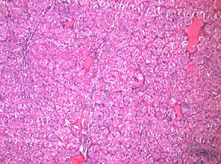
Figure 1: Acinic cell carcinoma 10x


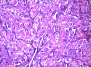
Figure 2: Acinic cell carcinoma 40x


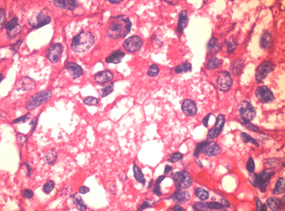
Figure 3: Acinic cell carcinoma 100x


Issue 2 Volume 12 - 2024