The analysis of the effect of etching of the carious cavity after preparation with a gel containing silver nanoparticles (Ag NPs) on the structure and strength properties of the "dentin- filling" and "enamel-filling" boundaries in permanent teeth with low caries resistance of hard tissues was carried out. The research samples were made from teeth removed for medical reasons in patients aged 18 to 60 years. The sealing was performed with adhesive systems of the IV and V generations, fluid and packable composites were selected as the filling material. Samples for metallographic studies were cut perpendicular to the main axis of the tooth, and their surfaces were polished mechanically and etched in concentrated orthophosphoric acid. The observations were carried out on light and scanning electron microscopes. It is shown that the etching gel does not lead to a decrease in the cohesive strength of the boundaries, despite the presence of silver there.
Introduction
The destruction of the adhesive compound "dentin-filling" and "enamel-filling" is considered one of the causes of the development of secondary caries, the prevention of which is an urgent task in dentistry [1-3]. One of the methods of relieving this pathology is remineralizing therapy, which allows to preserve the bioorganic phase in demineralized dentin [4, 5]. Therefore, the introduction of silver nanoparticles (Ag NPs) into the composition of dental materials due to its high bactericidal properties seems promising in preventing the development of secondary caries [6-8].
In modern medicine, Ag NPs find a very diverse application: wound treatment, surface disinfection, and implant coating [9-12]. The continuing interest in improving methods for producing Ag NPs is explained by the imperfection of existing technologies and attempts to improve such properties of the resulting product as stability and bioactivity [13, 14]. However, the specifics of the mechanism of Ag NPs influence on the adhesive compound "dentin-filling" and "enamel-filling" remain unclear (for example, whether silver-containing preparations will reduce the cohesive strength of the boundaries). When using such materials, silver infiltrates into the bioorganic matrix of demineralized dentin, which causes its remineralization [15-18]. The manipulation that ensures the delivery of silver to demineralized dentin and enamel is the etching of the prepared cavity [19, 20].
Thus, the aim of the study was to evaluate the effect of etching gel containing Ag NPs on the cohesive strength of the dentin-seal and enamel-seal boundaries in teeth with low caries resistance of hard tissues in mature individuals. Consequently, the main objective of the study is evaluation of the effect of etching of the prepared cavity with a gel containing silver nanoparticles on the strength properties of the "dentin-filling" and "enamel-filling" boundaries in permanent teeth of patients with low caries resistance of hard tooth tissues.
Materials and Methods
The study was performed on 46 teeth (premolars and molars removed for medical reasons in patients aged 40-60 years) with carious cavities of class I and II [21]. The prepared teeth were divided into two groups of 23 pieces each. The first group was a control group in which the teeth were treated according to the standard procedure (etching 15 c in 36% H3PO4) [22]. The second group was the observation group, where the teeth were treated for 15 seconds with Etchmaster Ag™ etching gel, which included 36% H3PO4 and a filler containing 10 ppm Ag NPs [12]. The prepared cavities in the teeth of both groups were filled with a fluid composite "Aeliteflo™", packable composites "Aelite All-Purpose Body™" and "Aelite Aesthetic Enamel™" using adhesive systems of the IV generation "All-Bond 3™" and V generation "Sealbond Ultima™". Postbonding of the surface of fillings and tooth enamel was performed with gels of the same types that were used for filling the control group and the observation group, and with Fortify™ sealant.
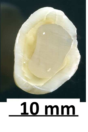
Samples for metallographic studies with a thickness of 1 mm were cut from the middle of the crown part of the tooth perpendicular to the main axis of the tooth (Figure 1). The surfaces of the samples were mechanically polished, after which the damaged layer was etched in concentrated H3PO4 for 2-5 minutes. The microstructure of dentin and enamel near the borders with the seal was studied using a metallographic microscope at magnification ×500. Studies at high magnifications were carried out on a scanning electron microscope JSM-6390LV [23]. The silver content in the tooth was determined using the LSX-500 laser ablation unit (Cetac) [12].
Results and Discussion
In the optical images obtained with an ×20 magnification, the enamel, dentin and filling differ in color, and their relative position in the crown is determined by the features of the filling installation in the tooth (Figure 1).
|
|
|
Figure 1. A sample cut from the crown of a tooth (×20) |
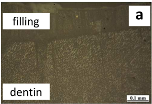
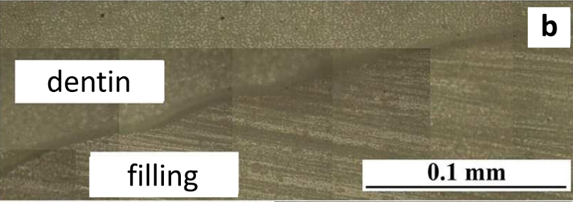
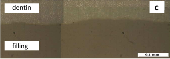
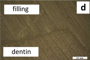
The boundaries between the hard tissues of the tooth and the filling are thin homogeneous lines that do not contain pores and cracks. There were no differences between the samples from the control group and the observation group. The observation data at an increase of ×500 confirm that the boundaries of the "dentin-filling" (Figure 2) and the "enamel-filling" are thin lines that do not contain defects. A slight etching of the boundaries, which was not noticeable with an increase in ×20, is due to the method of preparing the surface of the samples, which includes etching in concentrated orthophosphoric acid [24, 25]. Just as in the previous case, there were no differences between the boundaries of "dentin-filling" and "enamel-filling" in the samples of control and observation groups.
|
|
|
a) |
|
|
|
b) |
|
|
|
c) |
|
|
|
d) |
|
Figure 2. The "dentin-filling" boundary after treatment with an etching gel (optical microscope): a) with Ag NPs, adhesive system of the IV generation, b) Ag NPs, adhesive system of the V generation, c) without Ag NPs, adhesive system of the IV generation, d) without Ag NPs, adhesive system of the V generation |
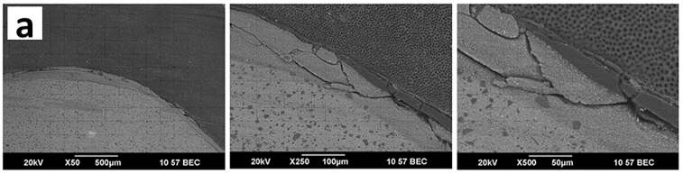
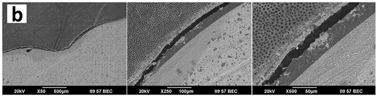
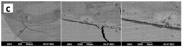
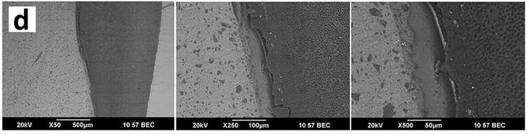
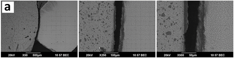
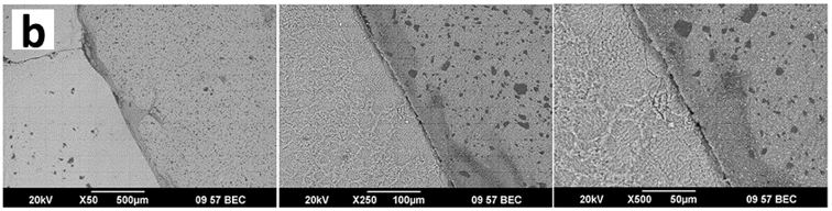
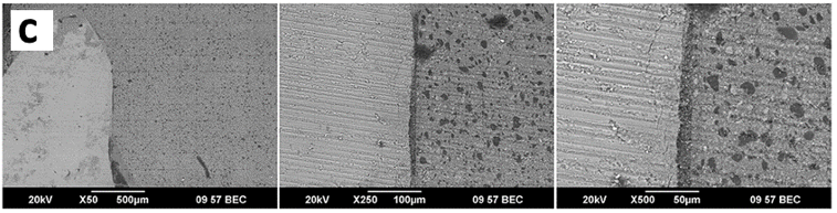
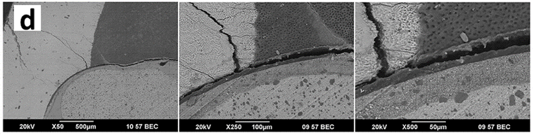
The results of the electron microscopic examination are consistent with the above data. The borders of "dentin-filling" (Figure 3) and "enamel-filling" (Figure 4) do not contain continuity defects and are evenly etched along the entire length. The appearance of a small number of microcracks at the boundaries is most likely a consequence of the mechanical impact exerted on the sample during tooth cutting and during mechanical polishing of the sample surface [26, 27]. In addition, the cracking of the boundaries can be affected by etching of samples in acid, as well as dehydration during storage, which can lead to deformation of the samples and destruction of the adhesive compound [28-30]. It should be noted that the growth of such cracks is inhibited in the surface layer of dentin or enamel, since the samples prepared for structural studies have never been destroyed along the boundaries, despite the fact that they were subjected to significant mechanical influences [31, 32]. Notably, electron microscopic examination of the boundaries did not reveal any differences between the samples prepared from the teeth of the control group and the observation group.
|
|
|
a) |
|
|
|
b) |
|
|
|
c) |
|
|
|
d) |
|
Figure 3. The "dentin-filling" boundary after treatment with an etching gel (scanning electron microscope): a) with Ag NPs, adhesive system of the IV generation, b) with Ag NPs, adhesive system of the V generation, c) without Ag NPs, adhesive system of the IV generation, d) without Ag NPs, adhesive system of the V generation |
|
|
|
a) |
|
|
|
b) |
|
|
|
c) |
|
|
|
d) |
|
Figure 4. The "enamel-filling" border after treatment with an etching gel (scanning electron microscope): a – with Ag NPs, adhesive system of the IV generation, b – with Ag NPs, adhesive system of the V generation, c – without Ag NPs, adhesive system of the IV generation, d – without Ag NPs, adhesive system of the V generation |
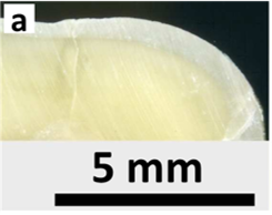
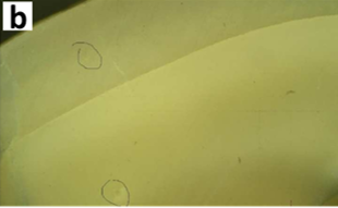
The study of the elemental composition of dentin and enamel of the control group samples, carried out at a distance of about 1.25 mm from the border with the filling, showed that a layer of hard tissue with a thickness of about 50 µm contains about 10ppm Ag (Figure 5). At the same time, silver was not found in the samples of the observation group.
|
|
|
a) |
|
|
|
b) |
|
Figure 5. Dentine-enamel compound treated with etching gel with Ag NPs: a) after cutting (there are no cracks or pores on the border), b) after studying the elemental composition in dentine and enamel (laser evaporation sites are circled) |
The use of physical materials science methods to study the cohesive strength of the "dentin-filling" and "enamel-filling" boundaries in the teeth of the control group and the observation group allowed us to draw two conclusions:
Conclusion
Thus, the results of the study showed that the presence of up to 10 ppm of Ag NPs in dentin and tooth enamel does not reduce the strength properties of restoration, therefore, the main function of silver in the etching gel can be considered a preventive effect on pathology.
It is worth noting that when using the results obtained in clinical practice, it should be borne in mind that the strength of the filling connection with the hard tissues of the patient's tooth depends on many factors, such as the clinical condition of dentin and enamel, the physico-mechanical properties of a particular filling material, the chemical properties of the adhesive system and the etching gel. Since in research, etching of dentin and enamel surfaces is carried out on grinds, which are prepared from teeth removed for medical reasons, this brings the working conditions as close as possible to natural ones. Based on the data obtained, it can be concluded that the main function of Ag NPs in the etching gel is a preventive effect on pathology, since its presence does not reduce the strength properties of restoration.
Acknowledgments: None
Conflict of interest: None
Financial support: None
Ethics statement: Teeth samples were obtained from patients after signing volunteer agreement for the use their biomaterial in the experiment. All raw data are available upon request from the corresponding author.