
INFLUENCE OF BACKGROUND AND CEMENT SHADES ON THE COLOR OF LOW-TRANSLUCENCY LITHIUM-DISILICATE CERAMICS
Mohammed Zahran 1*, Dania Sabbahi 2, Areej Abdulgader 3, Hanan Alrowithi 4, Mona Aqely 5, Alshaymaa Faydhi 6, Shahad Rushan 3
1 Oral and Maxillofacial Prosthodontics Department, Faculty of Dentistry, King Abdulaziz University, Jeddah, Saudi Arabia.
2 Dental Public Health Department, Faculty of Dentistry, King Abdulaziz University, Jeddah, Saudi Arabia .
3 Princess Nourah University, Riyadh, Saudi Arabia.
4 Private Practice, Jeddah, Saudi Arabia .
5 Al-Mikhwah Dental Center, Al-Baha, Saudi Arabia.
6 University Dental Hospital, King Abdulaziz University, Jeddah, Saudi Arabia.
|
|
INTRODUCTION
Metal-free dental ceramic materials were introduced into the field of dentistry by McLean since 1965. 1 Advancements in the ceramic industry improved our armamentarium with the introduction of ceramic restorations with optimal esthetics and mechanical properties. These advancements allow clinicians to meet their patients’ demands and expectations.2 However, dentists are often challenged with cases that involve matching single anterior tooth or implant-supported all-ceramic restorations to adjacent natural dentition, especially in the anterior zone. 3 This can be even more challenging when the case is involving a combination of natural dentition and implant-supported restorations as a result of different background colors. 4-7 Till recently, metal-based cement or screw-retained implant-supported restorations were the standard treatment option available for dental implants. Matching the color of metal-based to all-ceramic restorations can be challenging due to the dark color of the metal substructure. The introduction of strong ceramics, such as yttrium-stabilized zirconium oxide polycrystals (Y-TZP) materials allows a clinician to deliver implant-supported restorations with highly esthetic outcome and enhance the color matching to adjacent teeth. 8 These zirconia-based abutments are often combined with cement-retained all-ceramic crowns. Lithium disilicate is one of the most commonly used dental ceramics for this purpose due to their high mechanical properties and superior esthetics as a result of their optimal translucency which allows better light transmission thought the restoration. 9 Matching ceramic restorations made using these translucent ceramics with natural teeth is very challenging and cannot be ensured, 10 especially when cemented to natural teeth and/or implant-supported abutments with different stump shades.
Several factors might affect the final color of all-ceramic restoration. These factors can be divided into three main categories: ceramic-related, substrate-related, and cement-related. 11, 12 These factors include background color, luting cement color, and ceramic thickness. Having a dark background color (e.g. metal-based implant abutment, abutment teeth with intrinsic dark staining or metal cast post and core) would result in darker final restoration, and vice versa.6, 10, 13 Several studies evaluated the effect of luting cement shade on the final restoration color and concluded that using the opaquer shade of luting cement will help in masking the underlying dark substrate. 5, 7
Color matching in dentistry can be achieved either visually through the use of available shade guides (subjective method) or electronically by using instrument-based shade selection (objective method). Variety of dental shade matching devices have been introduced to overcome the limitations of the visual shade matching including, spectrophotometers, colorimeters, and imaging systems. Spectrophotometers are one of the most accurate and flexible instruments for color matching in dentistry. 13 They measure the energy of the transmitted or reflected light from an object at 1-25 nm intervals along the visible spectrum. 14 Compared to the subjective conventional method, spectrophotometers offered a 33% elevation in accuracy and a more objective match in 93.3% of cases. 14 The Commission Internationale de l'Eciairage (CIE) method was used frequently in the dental research to explain the relationship between color difference and their perception and acceptance in clinical practice. 15
The purpose of this in-vitro study is to determine the color difference using a variety of background shades and resin luting cement and their influence in approaching clinical reality. The null hypothesis was that there is no significant effect of the background shade and luting cement shade and their interaction on the color of low-translucent lithium disilicate glass-ceramic (LT-LDGC).
MATERIALS AND METHODS
All the materials used in the study and their specifications were listed in (Table 1).
Specimen Preparation
LT-LDGC blocks (IPS-e.max®CAD, Size C-14, Ivoclar/Vivadent, Liechtenstein) were sectioned to produce 60 ceramic rectangular specimens (15.75mm*14.25mm*1.5mm) utilizing diamond wafering blade (4”*0.012”) (UKAM Industrial Superhard Tools Valencia, USA) mounted on a slow-speed saw (Isomet 2000 Precision Saw, Buehler, USA).
Four different background groups were used to fabricate rectangle specimens (n=15/group). The specimens of the 1st (ZR) and 2nd (A3) groups were sectioned from zirconium-oxide blocks (IPS e.max ZirCAD, Size C15, Ivoclar/Vivadent) and composite blocks (Lava Ultimate, shade A3, 3M ESPE, Germany), respectively, using a diamond wafering blade (4”*0.012”) (UKAM Industrial Superhard Tools Valencia) mounted on a slow-speed saw (Isomet 2000 Precision Saw, Buehler). While, the specimens for the 3rd and 4th groups were fabricated from dual-cure composite core build-up material (LuxaCore Z-Dual, DMG, Germany) using light opaque (LO) and (A3) shades, respectively. The LuxaCore material was injected in a custom mold that was created using putty PVS material (Take 1 Advanced™ Putty, Kerr Corp., USA) utilizing lithium disilicate rectangular specimens (15.75mm*14.25mm*1.5mm). Then, cured using LED curing light (Bluephase N, Ivoclar/Vivadent) for a total of 40 seconds (20 sec/side).
All background and LT-LDGC specimens were polished using assorted silicon carbide sandpaper of different grits (220, 320, 400, and 600, sequentially) (ACE Hardware Corp., Oak Brook, IL, USA). After polishing, the specimens’ thicknesses (listed in Table 1) were verified using a digital caliper (Mitutoyo Corporation, Tokyo, Japan). ZR group specimens’ thicknesses were increased by 25% to accommodate for the anticipated shrinkage after sintering. LT-LDGC and ZR group specimens were crystalized/sintered according to manufacturer’s instructions using a porcelain furnace (Programat® EP 3010, Ivoclar/Vivadent) and a high-temperature furnace (Sintramat, Ivoclar Vivadent), respectively.
Cementation Procedures
The four background groups (A2, A3, LO, and ZR) were cemented to their corresponding LDGC specimens using self-adhesive dual-cure resin cement (Maxcem Elite Chroma, Kerr, U.S.A) with three different shades of cement; clear, white and yellow.
A standardized weight of 64g was applied over the ceramic/background complex during cementation To standardize the cement layer thickness for 5 min, followed by light curing using an LED light cure unit (Bluephase N, Ivoclar/Vivadent) for a total of 40 seconds (20 sec/side).
Spectrophotometric Measurement
A digital spectrophotometer (VITA Easyshade; VITA Zahnfabrik, Bad Säckingen, Germany) was used to record the color coordinates in the CIE (Commission International de l’Eclairage) Lab color space system. A custom-made square box of 6 cm3 lined with a black interior with an opening hole in the top of the box to accommodate the tip of the spectrophotometer during measurement.
Two spectrophotometric readings were recorded for each specimen, for ceramic specimens alone (S1) as a baseline and the ceramic-background complex after cementation (S2), respectively. All recorded data were organized and tabulated in an excel sheet (Microsoft Excel for Mac, version 16.10, 2017) to be prepared for statistical analysis.
To determine the effect of changing the background and the shade of the cement, color difference was calculated using the CIEDE2000 (DE00) according to the following formula:
∆E00=∆L'KLSL2+∆C'KCSC2+∆H'KHSH2+RT ∆C'KCSC∆H'KHSH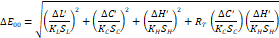
Where:
∆C′, ∆L′, and ∆H′: differences in chroma, lightness, and hue.
RT: rotation function which accounts for the interaction between chroma and hue differences within the blue region.
SL, SC, and SH: weighting functions that adjust the total color difference for variation in the location of the color difference pair in L, a, b coordinates.
KL, KC, and KH: parametric correction factors for experimental conditions.
The parametric factor of the CIEDE2000 color-difference formula was set to 1. While, the clinical acceptability threshold was set at DE00>2.25 units, and the perceptibility threshold was set at DE00=1.30. 16, 17
Statistical Analyses
Two-way analysis of variances (ANOVA), followed by Tukey’s post-hoc comparisons, was used to evaluate the effect of the background color, cement shade and their interaction on the color difference (DE00). All statistical analyses were two-tailed and conducted using the SPSS software for Windows (version 20, SPSS Inc., IBM, Somers, New York, USA) at a significance level of (0.05).
RESULTS
Two-way ANOVA revealed a significant effect of the background, but not for cement shade and the interaction term, on the color difference (Table 2).
|
Material and Manufacturer |
Lot No. |
Thickness (mm) |
Shade |
Type |
|
LuxaCore Z-Dual (LO) (DMG, Germany) |
763208 |
1.3 – 1.7 |
Light opaque |
Composite core build-up material (Barium glass in a Bis-GMA resin matrix) |
|
|
|
|
|
|
|
LuxaCore Z-Dual (A3) (DMG, Germany) |
779562 |
1.5 |
A3 |
|
|
|
|
|
|
|
|
IPS-e.max ZirCad (ZR) Ivoclar/vivadent, Liechtenstein |
L13763 |
1.5 |
White |
Yttrium-stabilized zirconium-oxide |
Table 1: Characteristics of the materials used in this study
|
Source |
Type III Sum of Squares |
df |
Mean Square |
F |
Sig. |
|
Corrected Model |
304.029 |
11 |
27.639 |
25.938 |
<0.001 |
|
Intercept |
1932.451 |
1 |
1932.451 |
1813.542 |
<0.001 |
|
Background |
297.300 |
3 |
99.100 |
93.002 |
<0.001 |
|
Cement |
3.037 |
2 |
1.518 |
1.425 |
0.251 |
|
Background * Cement |
3.692 |
6 |
.615 |
.577 |
0.746 |
|
Error |
51.147 |
48 |
1.066 |
||
|
Total |
2287.627 |
60 |
|||
|
Corrected Total |
355.176 |
59 |
Table 2: Two-way ANOVA (dependent variable: DE00; independent variables: background and cement shade).
R Squared = 0.856 (Adjusted R Squared = 0.823)
The mean color differences (DE00) of different groups in comparison to the baseline measurement are reported in (Table 3). The reported DE00 values ranged between 2 to 8.5. Most of the groups showed DE00 values higher than clinically acceptable level (DE00 >2.25). Only one group of the combination of ZR background and yellow cement showed mean DE00 value within the clinically acceptable level (1.3 < DE00 ≤ 2.25).
|
Background |
Cement Shade |
||
|
Clear |
White |
Yellow |
|
|
A2 |
7.9 (1.9) c |
7.3 (0.6) c |
7.1 (0.8) c |
|
A3 |
8.2 (1.0) c |
8.5 (0.8) c |
7.7 (1.6) c |
|
Luxacore (Lo) |
4.4 (0.8) b |
4.6 (0.7) b |
4.7 (1.3) b |
|
Zirconia |
2.7 (0.5) a, b |
3.0 (0.8) a, b |
2.0 (0.5) a |
Table 3: Mean (SD) for DE00 for different groups in the study (Results of Tukey post-hoc comparisons are shown as superscript letters, and values having the same superscript letters were not significantly different (P>.05))
DISCUSSION
The null hypothesis of the present study was partially rejected. The results revealed that the color perception of different background shades showed a statistically significant difference, while, the influence of cement shade and the interaction term were not statistically significant.
The structure of the lithium disilicate ceramic affects its optical properties. The presence of lithium disilicate crystalline phase dispersed in the glassy matrix allows the light to pass through ceramic without scattering, which might result in a reflection of the underlying background shade. 7 The findings of the present study confirmed that and revealed higher DE00 for the samples fabricated using A2 and A3 backgrounds as compared to the samples fabricated using ZR and LO backgrounds. This could be attributed to the white color of the ZR and LO backgrounds which reflect all the light that passes through the LT-LDGC compared to the darker backgrounds (A2 and A3) which reflect less and absorb more light. This finding is in agreement with previous studies. 5, 6, 18
In the present study, changing the cement shade did not mask the effect of the background color and did not help in tuning it significantly. This finding is in agreement with the results reported in a previous study. 4 On the contrary, a previous work that used a similar methodology reported a significant effect of the cement on the color difference. 18 This disagreement can be explained by the differences in the translucency of the glass-ceramic used in these studies. The present study used low-translucent in comparison to high-translucent glass-ceramic in the former study. LT-LDGC is expected to reflect more and pass less light to the underlying structure, which can mask the effect of the cement shade.
Three different shades of cement were used in the current study; yellow, clear, and white, which represents the range of shades available for dual-cure cement produced by different manufacturers. It is not clear whether using different cement brands might change the results of the study. This can be the main focus of future research.
In the present study, all the ceramic-cement-background combinations, except one, showed a high mean DE00 values that can be detected easily as clinically unacceptable (DE00 >2.5). The only combinations that revealed a clinically acceptable difference were zirconia background with yellow cement combined with LT-LDGC with DE00 = 2. This can be explained by the fact that yellow cement shade tuning of the highly reflective white background to match the color of the A3 ceramic. One of the other suggested solutions to overcome the high reflection of the white zirconia background is using different shades of zirconia or using an LDGC abutment attached to titanium base with a shade matching the all-ceramic crown.
Similar to low-translucent LDGC, the high-translucent version of LDGC failed to mask the color of the underlying background and cement combinations. 18 It seems that high and low translucent ceramics are not the material of choice in cases that involve abutments with a variety of stump shades since they have limited ability to mask the underlying substructure. 19, 20 Alternatively, using high opacity (HO) and medium opacity (MO) ceramic as a core structure veneered by veneering porcelain can be considered to provide better masking for the underlying background-cement combination and allow better shade matching. 7
In the current study, the clinically relevant ceramic thickness was selected as recommended by the manufacturer for all-ceramic lithium disilicate crowns. Increasing the ceramic thickness to 2.5 mm was found to improve color matching. 4 This increase might not be feasible in lots of situations and will contradict the concepts of conservative dentistry.
CONCLUSION
Within the limitation of this study, it seems that matching the color of adjacent LDGC restorations using LT ceramic is a difficult task. Varying the cement hue did not help to reduce the disparity in color between different substructure groups to a clinically acceptable level. Future research should focus on the effect of using opaque LDGC and different zirconia shades on the color matching.
REFERENCES
CORRESPONDING AUTHOR
Mohammed Zahran
Faculty of Dentistry, King Abdulaziz University, 3527 Abdullah Alsulaiman St.
Jeddah 22252
Saudi Arabia
Email: mzahran @ kau.edu.sa


