ANTERIOR TEETH VISIBILITY AND ITS RELATION TO AGING AMONG SAUDI POPULATION
Mohammad Abdullah AlRafee 1, Abdullah Saud AlRafee 2, Abdulaziz Saad Alanzan 3*, Hamad Saud AlRafie4, Abdullah Saleh AlGhureibi5, Mohammed Raja AlMujibah5, Razan Fahad AlEidan6, Reem Tariq AlDaijy5
1 Department of Prosthodontics , Dean College of Dentistry, Riyadh Elm University, Riyadh – 11681, Saudi Arabia.
2 Department of Restorative Dentistry, King Fahad Medical City, Riyadh – 11681, Saudi Arabia.
3 Dental department, Prince Mohammed bin Abdulaziz Hospital (MOH), Riyadh – 11681, Saudi Arabia.
4 Private Dental Practice, Alsafeer dental polyclinic, Riyadh – 11681, Saudi Arabia.
5Private Dental Practice, Riyadh – 11681, Saudi Arabia.
6 Department of Prosthodontics (KSMC), Riyadh – 11681, Saudi Arabia.
ABSTRACT
Aim: To examine the influence of age and gender on the degree of maxillary and mandibular anterior teeth display during natural and exaggerated smiles among a group of Saudi subjects.
Methods: 320 Saudi adults (males and females) were randomly chosen. The measurements were carried out directly on the subject at natural and exaggerated smiles utilizing a Fowler Electronic Digital Calliper to the nearest tenth of a millimeter. Three measurements per tooth were performd and the mean was determined. Data were analyzed utilizing SPSS.
Results: During both natural and exaggerated smiles, males demonstrated more of the maxillary right and left lateral incisors and canines and females displayed more of the maxillary central incisor. Conversely, males demonstrated more of the mandibular anterior teeth during natural smile and exaggerated smile than the females. With enhancing age, the amount of anterior teeth revealed during both natural and exaggerated smile reduced for the maxillary and enhanced for the mandibular teeth.
Conclusion: The degree of visibility of anterior teeth is measured by gender and its association with aging should be considered when providing aesthetic prosthodontics treatment.
Key words: Teeth visibility, anterior teeth, Esthetics.
Introduction
An attractive smile includes the harmonious interaction of the lip position 1-3, teeth 4, and related gingival architecture.5 The range of tooth display is measured by the lip line, as the lip moves vertically for the duration of smiling.6 Teeth visible during smiling are a noteworthy part of the anatomy of an esthetic smile.7 The 4 stages in a smile cycle are lips closed, resting display, natural smile, and expanded smile.8 Maxillary anterior teeth plays a crucial role in facial esthetics.9 The appearance of the anterior tooth surface with the lips throughout function is an essential factor in defining the outcome of operative dentistry, implant dentistry, the fixed or removable prosthesis, and orthognathic surgery.9
Dissimilarities in tooth display have been stated between subjects of diverse gender and age.10, 11 The identification of any imaginable association between tooth display, gender, and age is of interest as they could be utilized as a guiding principle to esthetic considerations in prosthetic restorations of maxillary teeth.12 Furthermore, age affects the amount of tooth visibility. With enhancing age, there is a lessened tooth display of the maxillary anterior teeth and enhanced display of mandibular anterior teeth.13 Henceforth, young subjects will show more maxillary than mandibular teeth, while the older subjects will reveal more mandibular rather than maxillary teeth.14
The exhibited length of anterior teeth can be one of the useful guiding principles for measuring the suitable vertical dimension of occlusion.15 It has been noted that, with the lips at rest, males reveal less maxillary central incisors than females. It was discovered that the mean vertical dimension of visible maxillary central incisors with the lips at rest was 3.40 mm and 1.91 mm in women and men respectively. Alternatively, it was 0.49 mm in women and 1.2 mm in men for the mandibular central incisors.14 Nevertheless, some populations revealed no association between dental morphology and gender.16
The degree of visibility of anterior teeth is measured by muscle positions that differ from one subject to another. In a research, males demonstrated more of the canine, maxillary lateral, and mandibular anterior teeth than females. Additionally, with enhancing age, the amount of maxillary anterior teeth that were visible at rest diminished.17 Another investigation described that, at rest and throughout smiling, maxillary tooth display lessened, and mandibular tooth display enhanced with increasing age. Though, the dissimilarities between age groups were not meaningful, except for maxillary central incisor display that reduced significantly as age enhanced.18
People with shorter upper lips exhibit more maxillary central incisor than those with longer upper lips. Then again, people with longer upper lip demonstrate more mandibular central incisors.14 It is of clinical interest to examine the impact of age and gender on the degree of tooth display in the maxillary and mandibular anterior region at the natural and exaggerated smiles as adequate data are missing at the present time. Therefore, this investigation was led to explore the impact of age and gender on the degree of maxillary and mandibular anterior teeth display throughout natural and exaggerated smiles among a group of Saudi individuals. This will help to determine the correct position of the anterior teeth in an edentulous arch.
Materials and Methods
This study was conducted over a period of 3 months from October to December 2017 at Riyadh Elm University and Public places in Riyadh City. 320 Saudi adults (males and females) were randomly selected. The subjects aged 18-60 years. All chosen individuals had maxillary and mandibular anterior teeth with no severe attrition, mobility, restorations, extrusion, caries, or any evident deformities. Subjects with a history of lip trauma, congenital abnormalities, oral and maxillofacial surgery, and individuals undergoing or had orthodontic treatment were excluded.
The measurements were carried out directly on the individuals at natural and at exaggerated smiles utilizing a Fowler Electronic Digital Calliper (Kevelaer, Germany) to the nearest tenth of a millimeter (Figures 1 and 2). The observable portion of the anterior teeth was determined vertically from the lip to the incisal edges of the incisor teeth and to the cusp tip of the canines at the mid-point of the tooth. The extent was considered to be zero when the tooth was not observed irrespective of how short it was. 3 measurements per tooth were performed and the mean was determined.
Statistical Package for Social Sciences (SPSS) Version 21 was utilized for the analyses. Descriptive analyses were carried out to present an overview of the discoveries. One-way analysis of variance (ANOVA) and independent samples t-test was carried out to measure statistically significant dissimilarities in mean values of the parameters (amount of teeth display, clinical crown length, and gingival display at rest and in the maximum smile). Alterations between different age groups for each group of teeth and related gingiva were carried out utilizing the Scheffe post hoc test. A p-value of ≤ 0.05 was considered as statistically significant.
|
|
|
(A) |
|
|
|
(B) |
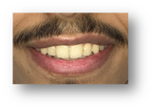
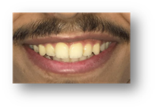
Figures 1. A: Natural smile, B: Exaggerated smile
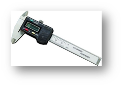
Figure 2. Electronic Digital Calliper
Results
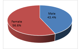
Figure 3. Distribution of participants by gender
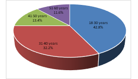
Figure 4. Distribution of participants by age group
Of the total 320 participants, most of them were females (56.6%) and between the age of 18-30 years (42.8%) (Figures 3 and 4). Most differences in the visible length of teeth surface during natural smiles were between the genders. The males exhibited more of the maxillary right and left lateral incisors and canines than the females. Nonetheless, the statistically significant difference between males and females was discovered only in the right and left lateral incisors (p<0.05). Oppositely, females exposed statistically significantly more of the maxillary central incisor than males (p<0.05) (Table 1). Males exhibited more of the mandibular anterior teeth during a natural smile than the females. Though, there was no statistically significant difference (p>0.05) (Table 2).
|
|
Mean±Standard Deviation |
|||||
|
Right |
Left |
|||||
|
Central incisor |
Lateral incisor |
Canine |
Central incisor |
Lateral incisor |
Canine |
|
|
Male |
2.32±1.92 |
1.94±1.01 |
0.97±0.80 |
2.42±1.62 |
1.89±1.11 |
0.89±0.43 |
|
Female |
2.98±1.45 |
1.25±1.32 |
0.56±0.34 |
2.84±1.59 |
1.65±1.03 |
0.61±0.31 |
|
p value |
0.000 |
0.035 |
0.062 |
0.001 |
0.029 |
0.084 |
Table 1. Mean amounts of visible maxillary teeth surface in a natural smile by gender (mm)
|
|
Mean±Standard Deviation |
|||||
|
Right |
Left |
|||||
|
Central incisor |
Lateral incisor |
Canine |
Central incisor |
Lateral incisor |
Canine |
|
|
Male |
1.31±1.01 |
1.24±0.93 |
0.71±0.58 |
1.33±1.09 |
1.25±1.11 |
0.78±0.67 |
|
Female |
1.10±1.05 |
1.03±0.84 |
0.69±0.42 |
1.19±1.10 |
1.04±1.01 |
0.70±0.51 |
|
p value |
0.081 |
0.723 |
0.421 |
0.605 |
0.108 |
0.092 |
Table 2. Mean amounts of visible mandibular teeth surface in a natural smile by gender (mm)
Likewise, males exhibited more of the maxillary right and left lateral incisors and canines for the duration of an exaggerated smile than the females. Nevertheless, a statistically significant difference was revealed only in the right and left lateral incisors (p<0.05). Females indicated statistically significantly more of the maxillary central incisor teeth than the males (p<0.05) (Table 3). Contrariwise, males showed more of mandibular anterior teeth during an exaggerated smile than the females. Though, there was no statistically significant dissimilarity (p>0.05) (Table 4).
|
|
Mean±Standard Deviation |
|||||
|
Right |
Left |
|||||
|
Central incisor |
Lateral incisor |
Canine |
Central incisor |
Lateral incisor |
Canine |
|
|
Male |
6.35±4.50 |
5.91±3.91 |
4.89±2.01 |
6.01±4.11 |
5.88±3.94 |
4.76±2.79 |
|
Female |
6.89±4.23 |
5.23±3.22 |
4.71±2.91 |
6.72±4.82 |
5.41±3.86 |
4.70±2.48 |
|
p value |
0.005 |
0.041 |
0.085 |
0.002 |
0.044 |
0.090 |
Table 3. Mean amounts of visible maxillary teeth surface in exaggerated smile by gender (mm)
With increasing age, the amount of maxillary anterior teeth that was visible during natural smile decreased. Among the anterior teeth, the amount of visible maxillary central incisors for the duration of natural smile was most significantly influenced by aging (p<0.05 (Table 5). With enhancing age, the amount of anterior teeth exhibited during natural smile enhanced for the mandibular teeth. Nevertheless, there was no statistically significant dissimilarity (p>0.05) (Table 6).
|
|
Mean±Standard Deviation |
|||||
|
Right |
Left |
|||||
|
Central incisor |
Lateral incisor |
Canine |
Central incisor |
Lateral incisor |
Canine |
|
|
Male |
3.85±2.16 |
3.01±2.11 |
2.83±1.83 |
3.99±2.64 |
3.25±2.56 |
2.71±1.46 |
|
Female |
3.42±2.71 |
2.89±2.22 |
2.62±1.79 |
3.42±2.70 |
2.75±2.04 |
2.55±1.91 |
|
p value |
0.812 |
0.081 |
0.621 |
0.492 |
0.077 |
0.102 |
Table 4. Mean amounts of visible mandibular teeth surface in an exaggerated smile by gender (mm)
|
Age group (Years) |
Mean±Standard Deviation |
|||||
|
Right |
Left |
|||||
|
Central incisor |
Lateral incisor |
Canine |
Central incisor |
Lateral incisor |
Canine |
|
|
18-30 |
3.72±2.19 |
2.11±1.54 |
1.78±0.91 |
3.88±2.45 |
2.21±1.15 |
1.62±0.75 |
|
31-40 |
3.11±2.01 |
1.73±1.10 |
1.13±0.72 |
3.33±2.60 |
1.66±1.32 |
1.10±0.56 |
|
41-50 |
2.74±1.76 |
1.11±0.77 |
0.76±0.21 |
2.81±1.85 |
1.03±0.63 |
0.66±0.31 |
|
51-60 |
1.63±0.89 |
0.93±0.43 |
0.45±0.39 |
1.72±0.69 |
0.89±0.44 |
0.30±0.25 |
|
p value |
0.030 |
0.077 |
0.741 |
0.043 |
0.058 |
0.213 |
Table 5. Mean amounts of visible maxillary teeth surface in a natural smile by age group (mm)
|
Age group (Years) |
Mean±Standard Deviation |
|||||
|
Right |
Left |
|||||
|
Central incisor |
Lateral incisor |
Canine |
Central incisor |
Lateral incisor |
Canine |
|
|
18-30 |
0.62±0.31 |
0.56±0.66 |
0.48±0.32 |
0.69±0.41 |
0.60±0.49 |
0.52±0.44 |
|
31-40 |
0.91±0.73 |
0.75±0.54 |
0.54±0.40 |
0.88±0.64 |
0.71±0.33 |
0.59±0.23 |
|
41-50 |
1.14±1.01 |
1.02±0.67 |
0.61±0.33 |
1.20±1.13 |
0.96±0.54 |
0.71±0.62 |
|
51-60 |
1.21±1.13 |
1.10±0.43 |
0.70±0.41 |
1.29±1.22 |
1.04±0.72 |
0.73±0.56 |
|
p value |
0.145 |
0.088 |
0.361 |
0.130 |
0.108 |
0.841 |
Table 6. Mean amounts of visible mandibular teeth surface in a natural smile by age group (mm)
With enhancing age, the amount of anterior teeth exposed during an exaggerated smile reduced for the maxillary and enhanced for the mandibular teeth. A statistically significant difference was revealed in the maxillary right and left central and lateral incisors (p<0.05). Contrariwise, no statistically significant difference was demonstrated in mandibular anterior teeth for the duration of an exaggerated smile (Tables 7 and 8).
|
Age group (Years) |
Mean±Standard Deviation |
|||||
|
Right |
Left |
|||||
|
Central incisor |
Lateral incisor |
Canine |
Central incisor |
Lateral incisor |
Canine |
|
|
18-30 |
6.77±4.32 |
5.71±3.57 |
4.41±2.23 |
6.85±4.56 |
5.63±3.40 |
4.36±2.17 |
|
31-40 |
6.24±4.11 |
5.10±3.05 |
4.10±2.03 |
6.51±4.32 |
5.04±3.12 |
4.18±2.11 |
|
41-50 |
5.44±3.67 |
4.59±2.88 |
3.66±1.63 |
5.52±3.71 |
4.46±2.34 |
3.44±1.30 |
|
51-60 |
4.62±2.50 |
4.01±2.42 |
2.94±1.02 |
4.70±2.44 |
3.93±2.11 |
2.75±1.10 |
|
p value |
0.010 |
0.030 |
0.070 |
0.003 |
0.046 |
0.611 |
Table 7. Mean amounts of visible maxillary teeth surface in an exaggerated smile by age group (mm)
|
Age group (Years) |
Mean±Standard Deviation |
|||||
|
Right |
Left |
|||||
|
Central incisor |
Lateral incisor |
Canine |
Central incisor |
Lateral incisor |
Canine |
|
|
18-30 |
1.62±0.92 |
1.10±0.65 |
0.94±0.49 |
1.98±1.03 |
1.13±0.82 |
1.01±0.37 |
|
31-40 |
2.13±1.82 |
1.86±0.82 |
1.21±1.03 |
2.08±1.12 |
1.94±0.75 |
1.61±1.15 |
|
41-50 |
3.35±2.13 |
2.74±1.91 |
2.10±1.41 |
3.22±2.05 |
2.81±2.03 |
2.25±1.52 |
|
51-60 |
3.88±2.42 |
3.11±2.27 |
2.44±1.69 |
3.80±2.31 |
3.44±2.65 |
2.69±1.73 |
|
p value |
0.413 |
0.104 |
0.800 |
0.514 |
0.419 |
0.734 |
Table 8. Mean amounts of visible mandibular teeth surface in an exaggerated smile by age group (mm)
Discussion
This investigation was carried out to examine the influence of age and gender on the degree of maxillary and mandibular anterior teeth display for the duration of natural and exaggerated smiles which will support defining the correct position of the anterior teeth in an edentulous arch. The visible portion of the anterior teeth has been unobserved by dentists as an element of esthetic evaluation. The amount of teeth display at rest which is acknowledged as the static position is mainly a muscle-determined position that differs from one subject to another.19 Quite the opposite, the dynamic position is normally characterized by a smile.20, 21
In the present study, the males displayed more of the maxillary anterior teeth than the females during a natural smile with the exception of the maxillary central incisor. In addition, males showed more of all the mandibular anterior teeth during a natural and exaggerated smile than the females. Previous studies reported that females displayed more maxillary incisor clinical crown length than males, with lips at rest during a maximum smile. 11, 22, 23 While some variations may be explained to some extent by differences in measuring techniques and differences between the populations studied. These findings were in agreement with the current study.
With the increasing age, the amount of maxillary anterior teeth exposed during natural and exaggerated smile decreased. However, the amount of mandibular teeth display enhanced with aging. These findings are revealed to be comparable to previous research.18 Facial aging is thought to be because of soft tissue variations among dentate patients.24 With the enhancing age, lips and the tissues surrounding the mouth sag have a tendency to be less elastic, leading to less maxillary tooth display and more visibility of mandibular anterior teeth.25 Additionally, a minor reduction could be attributed to attrition which lessens the clinical crown length of the teeth with age.26
One of the most beneficial guiding principles in defining the proper vertical dimension of occlusion is the tooth display of the anterior teeth.27 Additionally, esthetic considerations can be a major concern for patients looking for prosthodontic treatment.28 Concerning the amount of tooth display, it has been stated that the maxillary central incisor is a superior reference to the rest of the anterior teeth. Furthermore, it has been revealed that maxillary central incisors are the most dominant anterior teeth in the dental arch as they can be realized in their full size.9 Therefore, the maxillary right central incisor was utilized as a parameter to evaluate gender and age differences.29
The more the enhancement in the vertical dimension, the more display of the maxillary and mandibular teeth and vice versa. This general guideline will be more precise if the patient’s gender and age are considered as variables that may affect the visible amount of tooth at a natural and exaggerated smile. While restoring teeth, gender and age variances regarding the visibility of anterior teeth at natural and exaggerated smiles should be considered on a subject basis. The findings of the current study exhibit several variations between males and females, and different age group both at natural and exaggerated smiles.
Conclusion
The degree of visibility of anterior teeth is defined by gender and age. Males exhibited more tooth visibility of the maxillary lateral incisors and canines, and mandibular anterior teeth at natural and exaggerated smiles contrasted to female participants. Females exhibited more of their maxillary central incisor contrasted to males, both at natural and exaggerated smiles. With aging, the amount of anterior teeth displayed during an exaggerated smile reduced for the maxillary and enhanced for the mandibular teeth. These findings provide practical guiding principles for the vertical positioning of the anterior teeth
References
Corresponding Author
Abdulaziz Saad Alanzan
Dental department
Prince Mohammed bin Abdulaziz Hospital (MOH), Riyadh – 11681
Saudi Arabia
Email: Dralanzan @ gmail.com