RECOMBINANT AMELOGENIN PROTEIN ALONE REGENERATES LOST TISSUES IN IMMATURE TEETH WITH PULP NECROSIS AND PREAPICAL PERIODONTITIS
Maha Mohamed Fouad Mounir1, Ali Alharthi2, Jamaluddinsyed3, Madawi Alkeheli4*
1Oral Biology, Faculty of Dentistry, King Abdulaziz University, Jeddah, Saudi Arabia.
2Faculty of Dentistry, King Abdulaziz University, Jeddah, Saudi Arabia.
3Oral Basic and Clinical Science, Faculty of Dentistry, King Abdulaziz University, Jeddah, Saudi Arabia.
4Oral Diagnostic Science, Faculty of Dentistry, King Abdulaziz, Jeddah, Saudi Arabia. [email protected]
https://doi.org/10.51847/zPEhEt763f
ABSTRACT
In a research of immature apex closure carried out in 6 young mongrel dogs, polyglycolic acid (PGA) and amelogenin protein (RAP), generated by recombinant DNA technology, were compared.s. Pulp tissues of mandibular and maxillary premolars were completely removed during root canal treatment of all teeth. (n = 120). The cavities were left opened for 14 days. Canals were cleaned, watered, and divided equally for RAP or PGA treatment (n = 60). The treated teeth were removed from the mice after 1 and 3 months for histological analysis and immunological detection of protein markers linked to odontogenic cells. The animals were then killed. A pulp chamber with epulp-like tissue was found in amelogenin-treated canals after one month, and apical tissue that was functionally connected to the bone by a periodontal ligament PDL was also seen in amelogenin-treated canals analysed after three months. In contrast, although a calcified apical tissue was formed in the PGA group, neither soft connective tissue within the pulp chamber nor the periodontal ligament was observed. The findings suggest that recombinant amelogenin protein is the component that can signal for the lost dental tissue regeneration via activation of the Wnt signaling pathway and promoting cell survival and proliferation of neural stem/progenitor cells.
Key words: Amelogenin, Polyglycolic acid, Apex, Pulp, PDL, Wnt pathway.
Introduction
Pulp necrosis in immature secondary teeth with apical periodontitis is the most common in childhood because of trauma or caries. Extraction of these teeth at an early age may cause future problems such as loss of bone around teeth, migration of adjacent teeth, and occlusal discrepancies. Recently, regenerative endodontic treatment (RET), is an alternative to classical endodontic treatment for preservation of necrotic teeth with opened apex; it regenerates the pulp, restores the thickness of the dentinal walls, allowing for continuous development of the root and surrounding tissues, and restoring the root canals to a healthy state, thus regenerate sensory and defensive functions and restore functions of periodontal ligament and increase chances of survival of these teeth [1]. During clinical decision-making, the biodegradation In order to facilitate the new pulp/dentin matrix, the rate of the chosen biomaterial is an important component that needs to be taken into account in addition to its signalling potential and its capacity to distribute biomolecules in a sustained, controlled manner [2].
The total replacement of the original materials/scaffolds by the regenerating tissues is one of the most recent trends in pulp regeneration, and it is a sign of clinical success.. The objective of this study is to find the ultimate material that has the ability to regenerate lost dental tissue in immature teeth with apical periodontitis. For this purpose, we will check if Polyglycolic acid and recombinant amelogenin protein used alone are able to regenerate lost dental tissue in immature teeth with pulp necrosis and preapical periodontitis.
Pulp regeneration includes both the initiation of angiogenesis in order to improve nutrition (vascularization) and the sustainment of temp and pain sensitivity (innervation) and reconstruction and regeneration of the odontoblasts, the peripheral lining of the regenerated pulp, thus restoring not only the original tissue but also biotic functions. The dentinal pulp complex has a well organised histological structure, is vascularized, and contains a variety of cell types in distinct zones with a functional link to the surrounding dentin, making functional regeneration of the complex challenging [3]. Cell homing molecules are those that draw in the patient's own cells to promote tissue repair or regeneration. When excessive concentrations of endogenous stem cells are added into the wound area, making homing more clinically translatable [4]. Progenitor cell proliferation has the power to differentiate into all functional cell types and affect regenerative processes of the body [5].
One of the molecules that recruit the patient’s endogenous cells for cell homing is recombinant amelogenin protein (RAP) [6]. The essential enamel matrix protein is Amelogenin, which contains 90% of the enamel matrix in addition to 10% of different proteins, which include enamelin, amelins, and ameloblast [7].
Recombinant amelogenin protein (RAP) induces newly regenerated pulp-like cells in teeth with opened apex, resulting in the formation of calcified tissue in the apical foramen, which became connected to the bone via an orientated periodontal ligament in addition RAP induced the regeneration of pulp tissue similar to original pulp tissue in 85% in canals with nerves and nerve endings in the root canals of endodontically treated, non-vital immature teeth [7-9].
Most previous studies used RAP mixed with propylene glycol alginate PGA vehicle/scaffold to provide a body to the mixture [10]. Glycolic acid is a natural metabolite. It is a biodegradable polymer that loses its properties throughout approximately 2-4 weeks. It induces cellular migration and is assumed to be appropriate for neural regeneration and pulp tissue regeneration because of the adhesion and improvement of pulp fibroblasts. Additionally, it has an anti-inflammatory impact, which can be used for the remedy of endodontic damage [11-13].
An in vivo study was done in Japan comparing polyglycolide acid with fibrin glue bandage and gelatin sponge bandage and open wound in reconstruction after creating a three-dimensional defect in the skin and cranial periosteum of mice; post-operative wound healing was histologically assessed after one month, and a half the PGA group showed open wound; it showed an inhibition of number of osteoblasts and delayed healing [14].
Wnt/β-catenin signaling enhances Sox2 and Oct4 activity, which are core components of transcriptional networks that reinforce and regulate pluripotency. Wnt 1a, wnt 3a, wnt5, and Wnt 10b are detected in the regenerated tissues after amelogenin treatment in infected teeth with apical periodontitis [7, 10, 15].
Furthermore, Sox2 and Oct4 were also seen in the same tissues after amelogenin therapy. Sox2-Oct4 transcriptional networks are critical for the induction and reprogramming of somatic cells into induced pluripotent stem cells iPSC. Sox2 and Oct4 are able to induce a stable intrinsic pluripotency network ( in addition with environmental cues) that confers indefinite self-renewal capacity on iPSC [16-18].
The International Society of Cellular Therapy defined multipotent stromal cells as cells expressing CD antigens. Inflammatory cytokines and their receptors help regeneration and tissue repair [19].
CD74 is a cell surface receptor for the cytokine macrophage migration inhibitory factor (MIF), and its cell membrane receptor CD74 protects against injury and promotes repair and regeneration. Besides its critical role as a mediator upstreaming innate immunity, it also induces cell survival and proliferation of neural stem/progenitor cells NSPSs. MIF is a CD74 ligand that can activate various signaling pathways in cells. MIF is expressed endogenously in NSPCs to support the proliferation of Neural Stem Progenitor Cells NSPCs through CD74 ligand has the ability to activate many signaling pathways in cells [19, 20].
We aim to evaluate the ability of propylene glycol alginate alone/ and recombinant amelogenin protein alone to regenerate lost dental tissue in immature teeth with pulp necrosis and preapical periodontitis.
Materials and Methods
This study used a recombinant poly-histidine tagged murine 180 amino acid long Amelogenin (rp(H) M 180) that is identical to the authentic mouse full-length amelogenin. 30 mg of r-amelogenin was mixed with 1 ml of distilled water and was left for 15 minutes before use for group I. polyglycolic acid vehicle(Dexon)30 mg of polyglycolic acid (Dexon) was mixed with 1ml of saline for group II. Control canals left with no treatment. Six mixed-breed dogs aged 6 months were included in this investigation. One week prior to any endodontic treatments, animals were cared for and monitored for health assessments. In this investigation, 60 mandibular and maxillary premolars with 120 root canals each were employed. An intravenous injection of sodium pentobarbital (30 mg/kg body weight) was used to administer anesthesia. Prior to doing any operations, radiographs of the teeth's apices were taken to determine that all of the premolars had opened apices.
After gaining endodontic access, the length of each canal was measured, and the entire necrotic pulp tissue was removed using K-files in addition to Hedstrom files that were placed at the radiographic apex to guarantee full removal of all pulp tissue remnants. The surgical procedures were all carried out by the same surgeon.
The teeth were left without a coronal restoration for 14 days to allow the canals to become contaminated with oral microbes, and then the canals were cleaned to within 1 mm of the radiographic apices by the use of larger files as well as gentle filing motion with 1.5 percent sodium hypochlorite and irrigated with EDTA. Each canal was then dried with sterile paper points, and the teeth were sealed with a temporary filling (Orafil G, Colostol, Fermin, India). Seven days after closure, the temporary filling was removed, and the canals were filled using an angulated syringe with either Amelogenin (completely filling the canal) for half the specimens, group I n=30, and with PGA for the other half, group II n=30. Each procedure was followed by the administration of an intravenous painkiller and placement of intermediate restorative material (IRM) and sealing of the access cavity. Voltaren (Novartis Pharma Egypt, under license from Novartis Pharma, Switzerland), and 25mg/kg. On the first day, amoxicillin (Cid Co, Egypt) was given intramuscularly. The following seven days, a dose of 15 mg/kg of amoxicillin was mixed with food. To lessen the chance of local trauma to the treated teeth during the recovery period, the dogs were given a soft food. The animals were put to death by an intravenous thiopental sodium overdose injection one to three months after surgery. Using a water-cooled diamond disc, the teeth and surrounding bone were removed as a block, and samples were examined. To acquire roughly 60 canals at each passing time interval, the treated teeth were recovered at 1 and 3 months after treatment. Samples were demineralized, and 5 m-thick tissue slices were prepared using customary histologic techniques.
The thickness that was stained using hematoxylin and eosin and for immune detection of induced pluripotent stem cells (iPSC) and/or neural stem/progenitor cells NSPS by recombinant Anti-CD74 antibody [EPR25399-94] Eliza study: Mouse Cd74 (HLA class II histocompatibility antigen gamma chain) ELISA Kit Catalogue Code: MOFI01321.
For preparation, Tissue Homogenates: Tissue is rinsed with 1X PBS to remove excess blood, then the tissue is minced after weighing and homogenizing in PBS (the volume depends on the weight of the tissue. 9 mL PBS (including protease inhibitors) would be appropriate for 1 gram of tissue. For further lysing cells, the suspension is sonicated with an ultrasonic cell disrupter, or it is subject to freeze-thaw cycles. The homogenates are then centrifuged for 5 minutes at 5000 x g, and the supernatant is removed for assaying. The total protein concentration was determined by the BCA kit( the total protein concentration of each pore sample should not exceed 0.3mg). Sandwich Elisa has been used for the detection of antigen-induced Pluripotent stem cells.
Step 1: Capture antibodies specific to the target antigen that are immobilized in a microtiter well.
Step 2: Well is rinsed to remove unbound antibodies.
Step 3: Sample (containing the Ag) is added.
Step 4: Well is rinsed to remove unbound and non-specific antigens.
Step 5: Goat anti-rabbit IGg (H+L) Secondary Antibodies are added to the well.
Step 6: Well is rinsed to remove the unbound Secondary Antibody.
Step 7: Substrate specific for Enzyme-linked antibodies is added
Step 8: Color development indicates a positive reaction and present of antigen in the test sample, measure it through microplate reader.
Statistical analysis
Data collected were analyzed using SPSS program version 26.0. One way ANOVA test was used to examine the difference between the three study groups regarding the testing of levels and quantitative results of CD74 in tissue homogenates (Supplemental Tables).
Results and Discussion
This study was performed comparing CD74 antibody reactions between three groups, control, Amelogenin, and PGA, at three different times, 3,7,15 days by using a one-way ANOVA test and revealed the outcomes were significantly different, with a P-value <0.05 (Table 1).
Table 1. Eliza of control, polyglycolic acid, Amelogenin in three different time intervals: 3,7,15 days.Amel= Amelogenin, PGA= Polyglycolic acid, df= degree of freedom, F= F-value, S= Significance
|
|
Sum of Squares |
df |
Mean Square |
F |
Sig. |
|
|
Control |
Between Groups |
.021 |
2 |
.010 |
43.570 |
.000 |
|
Within Groups |
.004 |
18 |
.000 |
|
|
|
|
Total |
.025 |
20 |
|
|
|
|
|
amlg |
Between Groups |
22.340 |
2 |
11.170 |
17.659 |
.000 |
|
Within Groups |
11.386 |
18 |
.633 |
|
|
|
|
Total |
33.726 |
20 |
|
|
|
|
|
PGA |
Between Groups |
.487 |
2 |
.243 |
1.694 |
.002 |
|
Within Groups |
2.586 |
18 |
.144 |
|
|
|
|
Total |
3.072 |
20 |
|
|
|
|
Regenerative study results
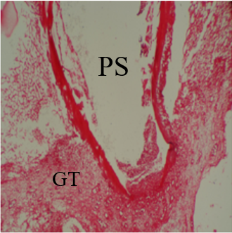
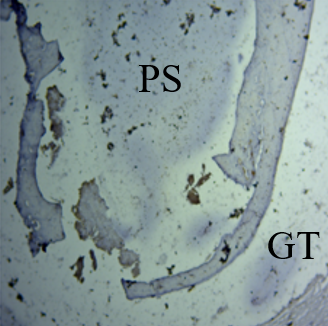
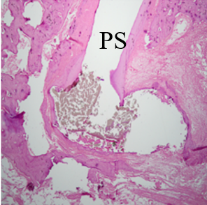
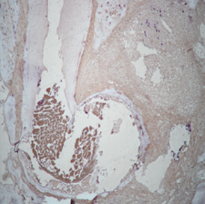
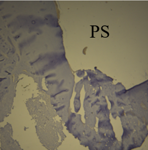
One month: The root apex is closed or partially closed in 10% of specimens only. The root apex is surrounded by granulation tissue in 20% of specimens. The pulp space is empty (Figure 1a). The root apex is not surrounded by granulation tissue in 80% of specimens and shows no IR to CD 74 in either granulation tissue or empty root canals (Figure 1e).
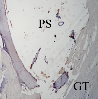
Three months: Root apex shows abscess formation in 40% of specimens (Figures 2a and 2e).
PGA-treated canal cohort
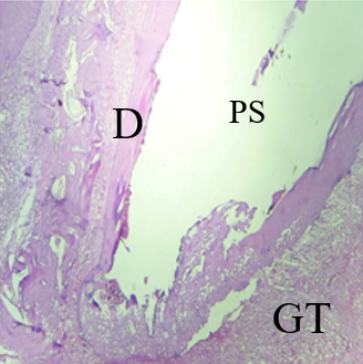
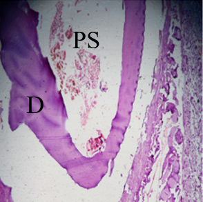
One month: As the root apices expand, granulation tissue made of cellular proliferation can be seen surrounding them. (Figure 1b). Apices show 45% root closure (Figure 1b), and 55% of roots are still opened (Figure 1f). The cells of the granulation tissue/dental follicle show no IR to CD74 proteins (Figure 1f). Pulp space is empty.
Three months: Shows complete root apex closure in 75% of specimens, and 25% still open and incompletely closed apex. The root apex in 40% of specimens is not closely surrounded by granulation or dental tissues. Regenerated bone is seen near the closing apex. The pulp space is empty or shows some debris (Figure 2b). Neither the closed root apex nor the surrounding granulation tissue shows IR to CD 74 (Figure 2f).
Amelogenin-treated canal cohort
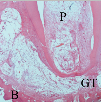
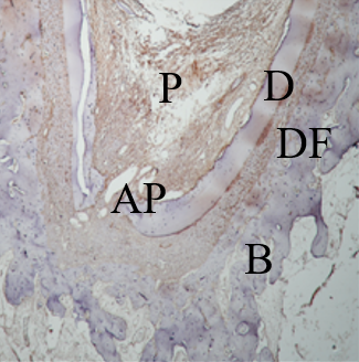
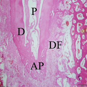
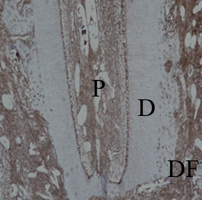
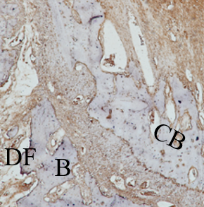
One month: Shows delicate vascular cellular proliferation surrounding the growing root apices (Figures 1c and 1d). Apices show 75% root closure (Figure 1c), and 25% of roots are still opened (Figure 1d). The cells of the dental follicle/dental papilla show moderate to intense IR to CD74 proteins (Figures 1g and 1h). The regenerated vascular and cellular pulp tissue fills the once-empty root canals (Figures 1c and 1d). It shows IR to CD74 proteins (Figures 1g and 1h). The odontoblasts in the regenerated pulp show IR CD74 (Figures 1g and 1h).
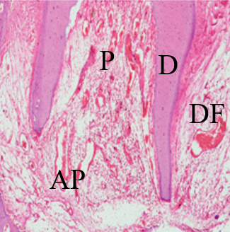
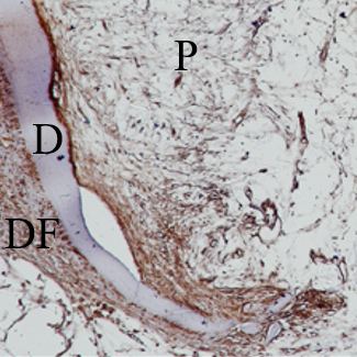
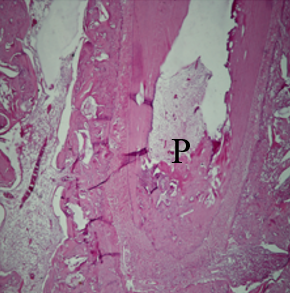
Three months: The root apex is closed in nearly 100% of specimens. Roots are surrounded by developing cementum, periodontal ligament PDL, and bone. Root canals are filled with regenerated delicate tissue representing the developing pulp (Figures 2c and 2d). The regenerated cementum, PDL, and bone show moderate-intense IR to CD 74 proteins. Also, the regenerated pulp tissue and odontoblasts show moderate IR to CD 74 (Figures 2g and 2h).
|
|
|
|
a) |
b) |
|
|
|
|
c) |
d) |
|
|
|
|
e) |
f) |
|
|
|
|
g) |
h) |
|
Figure 1. Premolar teeth at selected one-month time intervals following treatment. |
|
|
|
|
|
a) |
b) |
|
|
|
|
c) |
d) |
|
|
|
|
e) |
f) |
|
|
|
|
g) |
h) |
|
Figure 2. Premolar teeth are selected at a 3-month time interval following treatment |
|
This study defines the materials used for dental tissue regeneration, which includes dentin, cementum, periodontal ligament, and alveolar bone, that have complicated requirements in their mechanical properties, bioactivity, and necessities of carrying out many functions. Numerous proteins for clinical utilization have been studied, including amelogenin protein and polyglycolic acid PGA.
The objective of our study is to find the ultimate material that can regenerate lost dental tissue in immature teeth with apical periodontitis. For this purpose, we will check if Propylene glycol alginate and recombinant amelogenin protein are able to regenerate lost dental tissue in immature teeth with pulp necrosis and preapical periodontitis, each material alone or combined.
Apical periodontitis results from the pathogenic effects of the micro-organisms colonizing the root canal system and the response of the host defense system against this invasion of the periradicular tissues. Infected periradicular tissues showed varied severity of tissue destruction, possibly due to the virulence and number of pathogenic micro-organisms that invaded the canal.
Root canal treatment (RCT) is the most common treatment method for apical periodontitis. However, the complete removal of pulp tissue means loss of a warning system, immune response, and dentin formation as active defense mechanisms against invading toxins and bacteria. Regenerative endodontic treatment is then the treatment of choice; both materials, Amelogenin and PGA, are biocompatible [8, 9]. Amelogenin has an anti-inflammatory impact [21]; this impact is revealed in the destruction of the invading micro-organisms, resulting in the repair and regeneration of the periradicular tissues. On the other hand, PGA is said to induce local inflammation [22]. Only the amelogenin group reveals the closing of the opened apex by dentin regeneration, cementum, and periodontal ligament regeneration in addition to pulp regeneration. The PGA group shows only the closure of the opened apex by regenerated dentin. In contrast, the peri-radicular tissues show destruction, and only in the 3-time period it shows granulation tissue. The control group shows the persistence of inflammation and infection.
In a variety of experimental settings, regeneration of pulp-like tissue has been demonstrated [23]. Activation of stem cells, in vivo cell mobilization, and homing-inducing endogenous reparative cells are key components of the field of regenerative endodontics. The regenerative powers of RAP may be due to the utilization of the Canonical Wnt/β-catenin signaling. The amelogenin signaling cascade activates Wnt/β-catenin signaling that increases nuclear and cytoplasmic β-catenin. Increased β-catenin initiates DNA replication and cell proliferation and growth of stem/ progenitor cells and neural stem/progenitor cells NSPSs [7]. All regenerated tissues that are seen post amelogenin therapy revealed the expression of Wnt family members [19].
[24] Following amelogenin therapy, all tissues that had been regenerated showed the expression of Wnt family members. The Wnt/-catenin pathway is self-reinforcing once it is engaged. This could explain the amelogenin protein's special capacity for regeneration [17, 25, 26].
Damaged tissues achieve regeneration mainly through triggering an inflammatory response that protects the host and activates the regeneration process, in addition to the application of stem cell homing approaches. The vacant space of the infected debrided root canal is either empty or contains tissue remnants and debris. Periapical activation by key molecules that can restore the molecular machinery for regeneration is required to restore the destructed tissues and populate the vacant root canal space with an innervated tissue supported by vascularity.
Recombinant Amelogenin in regenerative endodontics regulates stem cell pluripotency via Wnt/β-catenin by inducing the expression of Sox2 and Oct4 pluripotency transcription factors in the regenerated periapical tissues and the lost dental pulp. Sox2 accelerates healing and derives induction and self-renewal of pluripotent cells after a tooth is injured or subjected to caries [27-29].
Induced pluripotent stem cells iPSC induction by Wnt/β-catenin signaling through the expression of Sox2 pluripotency transcription factor results in differentiation into diverse cell types and tissues, restoring their pluripotency state.
Canals treated with PGA showed no reactivity to ELISA or immune reactivity to CD74 in both periods.
The histological findings in the one-month post-operative period show the re-appearance of the embryonic tissue of the apical papilla and dental follicle that fills the periapical area at the injury site. The apical debrided and the previously empty root canal show aggregation of stem cells immune reactive to the amelogenin group, indicating the induction of iPSCs and embryonic tissues of the apical papilla, dental follicle, and immature pulp tissue that were present in this area during tooth development. In the three-month post-operative period, the amelogenin-treated canals show the presence of vascularized, innervated pulp-like tissue containing stem cells immune reactive to CD 74. Wnt/β-catenin signaling has the ability to regulate pluripotency. The endogenous stem cells result in tremendous regenerative capacity by inducing pluripotent stem cells [16, 30, 31].
Treatment with Amelogenin without the addition of PGA also induced pulp regeneration immuno-r the presence of neural elements in pluripotent t induction of pluripotent stem cells and the presence of neural elements he active to CD74 denoting PGA synthetic polymer is often used as a scaffold for tissue regeneration and cell seeding. Synthetic polymers' fundamental drawback is their inability to deliver the biochemical signals required to "communicate" with cells. Synthetic polymers can be functionalized by including one or more signaling molecules, such as amelogenin peptide, to get around this problem. Histological results of the PGA group show some regenerative effects, such as root dentin closure, but complete regeneration of the attachment apparatus and dental pulp tissue was not recognized. In addition, to the amelogenin peptide, the regeneration was upgraded, and complete regeneration of dentin, pulp, and tooth attachment apparatus was seen.
PGA was reported to inhibit osteoblast and delay healing in wounds and bone defects [13, 14, 32].
Recently, the most promising technique for preserving teeth with immature apex is regenerative endodontics, which restores the root canals to a healthy state, allowing for continued development of the root and surrounding tissues with increased thickness of dentinal walls [3]; during clinical decision-making, the most important factor to be considered. The complete replacement of the initial materials/scaffolds by the regenerating tissues is an indication of clinical success. Another important factor is that the introduced biomaterial is be able to deliver active biomolecules in a sustained pattern to facilitate the deposition of new pulp/dentine matrix [23].
Conclusion
Clinicians may use both materials in regenerative endodontic procedures to gain regeneration of all the lost dental tissues due to necrosis and periodontitis.
Acknowledgments: None
Conflict of interest: None
Financial support: None
Ethics statement: None