EFFECT OF VARIOUS ANTIBACTERIAL MATERIALS IN DENTAL COMPOSITES: A SYSTEMATIC REVIEW
Badr Soliman Alhussain1, Abdulrahman Abdulaziz Alfayez2*, Abdullah Abdulrahman Alduhaymi2, Essam Abdulaziz Almulhim2, Mohammad Yahya Assiri2, Shahzeb Hasan Ansari3
1 Department of Consultant Restorative, PSMMC, Riyadh, KSA.
2 Department of Dentistry, Prince Mohammed bin Abdulaziz Hospital, Riyadh, KSA. [email protected]
3 Department of Faculty Preventive, Faculty of Dentistry, Riyadh Elm University, Riyadh, KSA.
ABSTRACT
Dental composites are used in the treatment of conditions such as caries but could also produce marginal fractures and the emergence of secondary dental caries. The purpose of this study is to investigate the effects of antibacterial agents used with contemporary composites in managing dental caries. The systematic review involved the identification of relevant articles on the Google Scholar, Cochrane Library, and PubMed databases. Studies published between 2011 and 2021 were obtained, screened, and assessed for eligibility using the PRISMA guidelines. The review resulted in an analysis of 10 studies that adopted experimental and in-vitro or in-vivo investigation approaches to propose new materials or investigate existing materials’ antibacterial, mechanical, and aesthetic impacts on the composites and dental structures. The studies revealed that the antibacterial agents are effective in inhibiting the growth of caries-causing bacteria or killing the bacteria. The materials also had regenerative, remineralization, and protein-repellent effects on the dental structures and the composites. The poor material design could, however, affect the mechanical properties of the composites and the teeth. Novel antibacterial agents used in dental composites have a positive impact on the prevention of dental caries, restoration of lost tooth minerals, regeneration of the dental structure, and the repulsion of proteins.
Key words: Antibacterial, Agents, Composites, Dental, Mechanical, Aesthetic.
Introduction
Dental injuries or diseases can produce tooth caries, trauma, and other complications that result in the loss of the tooth structure. In the treatment of structural damages to the teeth, various materials have been used over the years, with synthetic materials being the major focus in dentistry since the early 19th century [1-3]. Their limitation, however, is their inability to replace and restore the structures of the tissues [1]. Consequently, other materials have taken center stage in dental practice and have drawn the interests of researchers and dentists alike. Zheng et al. (2019) present resin-based composites as one of the materials that have found use in restoring the structure and cementing crowns and veneers in place of dental amalgam, which is slowly phasing out in practice.
Of all the materials used in the management of dental structural damages, resin-based composites have received the most attention. Aminoroaya et al. (2021) highlight how these materials have become promising for tooth resembling in the practice of restorative dentistry [4]. The major limitation that this type of composite has is the occurrence of bulk or marginal fractures and the development of secondary caries when they are used, which limits the longevity of their restorative capabilities [4]. For instance, Kasraei et al. (2014) mention that composite resins that are manufactured using silver and zinc oxide have no antibacterial properties, with the two chemicals being broad-spectrum antimicrobials [5]. Thus, their use also increases the risk of developing secondary caries [5]. In the management of the emergent secondary caries with the use of resin-based composites, other researchers and dentists have explored the use of antibacterial fillers in composites. For instance, Stencel et al. (2018) explored the use of silver sodium hydrogen zirconium phosphate (SSHZP) antibacterial filler in the prevention of the survival of cariogenic bacteria arising from the use of dental composites in structural restorations [6]. The filler was beneficial to the reduction in the bacteria found in the teeth after the use of composites.
Similarly, Sun et al. (2021) presented the case for the use of a new generation of antimicrobial dental polymers to check the development of secondary caries and to elongate the life of the restorations done through resin-based composites [7]. The effects of the antimicrobial materials include inhibiting the formation of biofilms in the dental structures, reducing the rate of production of acids by the bacteria present in the teeth after the use of composites, and eliminating the occurrence of caries [7]. Korkut et al. (2016) demonstrated the efficacy of using bioactive glass in dental resin composites to inhibit Escherichia coli, Staphylococcus aureus, and Streptococcus mutans bacteria [8]. Chen et al. (2018) present a review of various materials that have been found useful as antibacterial agents for experimental and commercial production of dental restorative materials [9]. The agents that they highlight include leachable compounds, monomers that can be polymerized, and filler particles such as silver nanoparticles.
Other researchers have also revealed how new composites prevent the growth of bacterial colonies and biofilms, inhibit acid production, re-mineralize the teeth, and help in the healing of cracks. Yao et al. (2020) highlighted these positive impacts of chlorhexidine (CHX), silver, and fluoride that are used in dental polymers, the adhesive effects of antibacterial resins, the use of fluoride ions to enhance re-mineralization, and self-healing effects of polymers such as capsule-based, vascular, and intrinsic healing systems [10]. According to Villegas et al. (2019), resins loaded with zinc nanoparticles have re-mineralization action when used on the demineralized surface, and also have an improved dental power that makes them suitable for use in caries lesions [11]. The purpose of this study is to investigate the effects of the antibacterial agents used in dental composites on the properties of the resulting materials.
Materials and Methods
Search strategy
The process of identifying the articles that address the topic of study involved the conduction of an online search on the Google Scholar, Cochrane Library, and PubMed databases. The search was performed on July 7, 2021, using “dental composites,” “antibacterial materials in dental composites,” and “effects of antibacterial materials in dental composites” as the keywords. The researcher reviewed the titles of the articles and their abstracts for potential inclusion in the study. Only the articles whose titles and abstracts matched the purpose of the study were considered for the eligibility analysis. Further, the relevant studies that were cited by the authors of the considered articles were also analyzed for eligibility.
Study eligibility
From the literature search and identification of relevant articles based on the titles and abstracts, the researcher then conducted an eligibility analysis of the articles. The criteria used to examine whether the articles could be included in the systematic review were as follows:
Based on the criteria, various articles with the right information and relevance to the topic of the study were considered for the systematic review. However, to further narrow down the number of articles that could produce a high-quality systematic review, three exclusion criteria were applied in further eligibility studies. They were as follows:
Data extraction and analysis
After the eligibility analysis, the full manuscripts of the articles that met the inclusion criteria and did not meet the exclusion criteria were retrieved, with the researcher determining the final inclusion. The Cochrane Risk of Bias Tool was used to assess the extent to which design flaws, analysis and conduct procedures, and the reporting of the studies – especially the randomized clinical trials – resulted in the overestimation or underestimation of the impacts of the materials studied [12]. The final list of articles from the process was then used to extract data that provide evidence for conclusive findings on the effects that antibacterial materials used in dental composites have. The information was then summarized thematically based on the purpose of the study. The summary also included the risk of bias associated with the specific study.
Results and Discussion
Study selection
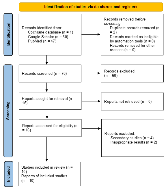
The initial literature search yielded a total of 78 articles with relevant titles, with 47 from PubMed, 30 from Google Scholar, and one (1) from the Cochrane database. Two of the articles identified were duplicates, and 76 others were then screened. From the use of the criteria specified above, 60 articles were excluded from the study, and 16 were assessed for eligibility. Four more articles were excluded from the study because they were secondary sources – systematic and literature reviews – and two others were excluded because of the inappropriateness of the results to the study topic. The PRISMA flow chart in Figure 1 details this process.
|
|
|
Figure 1. Study identification chart (PRISMA) |
Study characteristics
The included studies were based on experimental, in-vitro, and in-vivo methods of investigating the actions of the materials as part of dental composites. The maximum number of antibacterial agents investigated as part of the composites was three, with each study considering at least one agent. The focus of the studies was on the efficacy of the materials in inhibiting bacterial growth, killing bacteria, and effects on the properties of the composites used in dental treatments. None of the studies involved the trial of the produced materials on a patient since they were mostly experimental.
Study findings
Table 1 specifies the approaches that the authors used in their studies, the number of materials they investigated, the conclusions they arrived at, and the Cochrane Risk of Bias Tool determination.
Table 1. Study overview
|
Authors |
Design, year published |
Material number |
Findings |
Risk of Bias |
|
Hollanders et al. |
In vitro study of restored enamel-dentine blocks, 2020. |
30 blocks, using 3 materials |
The effectiveness of antibacterial bonding materials reduces over time. Tested materials included conventional bonding composites, antibacterial bonding composites, and amalgam. Lesions get deeper in the blocks with antibacterial bonding as the age of the blocks. |
Other bias: Authors do not describe the uptake of proposed materials.
Risk of bias: moderate. |
|
Hegde et al. |
Experimental in-vitro study with quantitative statistical testing of control and restorative materials, 2018. |
3 materials |
Nano-hybrid composites, glass ionomer cement (GIC), and silver amalgam showed an inhibitory effect against a Streptococcus bacterium. The silver amalgam produces the strongest inhibitory effect. |
Selection bias: Authors do not provide criteria for separating materials into test and control groups. Risk of bias: low. |
|
Peralta et al. |
Experimental evaluation of mechanical and physical properties of resin-based materials with quantitative statistical analysis, 2018. |
2 materials |
Some antibacterial materials in composites – Fermit inlay – inhibit the accumulation of Streptococcus mutans biofilms, while others – Luxatemp LC and Bioplic – have a continuous effect against the Enterococcus faecalis bacteria. |
Other bias: Authors do not describe the uptake of proposed materials.
Risk of bias: low. |
|
Chatzistavrou et al. |
Experimental evaluation and characterization of designed dental novel materials used in composites, 2014. |
1 material – glass ceramics |
The incorporation of silver ions in bioactive glass-ceramics used in dental applications produces a stable material that kills bacteria. The resulting material can also be used in tooth regeneration processes. |
Other bias: Authors do not describe the uptake of proposed materials.
Risk of bias: low. |
|
Bariker & Mandroli |
Experimental, using the agar diffusion technique to assess, 2016. |
2 materials |
Both materials – Amalgomer CR and Fuji VII – had antibacterial action against the variety of microorganisms that cause severe childhood dental caries. |
Other bias: Authors do not describe the uptake of proposed materials. Risk of bias: low. |
|
Park et al. |
Experimental, mixing MPC and MBN in different ratios with orthodontic bonding agents, assessing antibacterial and remineralization effects, 2020. |
2 materials – MPC and MBN, mixed with bonding agents |
The synergy of the bonding agents increases with the addition of MPC and MBN at the appropriate ratios, with the effects being antibacterial, protein-repellent, and anti-demineralization. |
Other bias: The authors do not provide a rationale for selecting the two materials. Risk of bias: low. |
|
Yaghmoor et al. |
Statistical analysis using ANOVA and pairwise comparisons of novel antibacterial composites, 2020. |
Composite with 2 materials – polylysine and monocalcium phosphate monohydrate |
A controlled release of polylysine in gaps caused by caries kills the bacteria and has a positive effect on the prevention of recurrent caries. |
Selection bias: Authors do not provide criteria for separating materials into test and control groups. Risk of bias: low. |
|
Yang et al. |
Development of complex antibacterial agents in dental composites, 2021. |
1 composite used |
The introduction of zinc oxide particles to nanoparticle composites does not corrupt their regular shape and close-packed structure. |
Other bias: The authors do not provide a rationale for rejecting other materials. Risk of bias: low. |
|
Al-Dulaijan et al. |
Synthesis of composites, experimental measurement of their ion release and recharge properties, 2018. |
2 composite materials were produced and tested |
The flexural strength and elastic modulus of the produced composites were commercially viable. The materials limited the growth and colony formation of biofilms. |
Other bias: The authors do not provide a rationale for rejecting other materials. Risk of bias: low. |
|
Zhang et al. |
Experimental design and testing of a new method of producing antibacterial composites, 2014 |
1 antibacterial agent used |
Dental composites containing chlorhexidine entrapped in mesoporous silica nanoparticles had a positive effect on the mechanical properties of the filler material. |
Other bias: The authors do not provide a rationale for rejecting other methods. Risk of bias: low. |
The review of ten (10) articles investigated the effects that various antibacterial materials in dental composites have, revealing both the negative and positive impacts. The antibacterial materials used in dental restoration inhibit the growth and multiplication of some bacteria. Hegde et al. (2018), after testing the impacts of a nano-hybrid composite, GIC, and silver amalgam against Streptococcus mutans, report that all the restorative materials have an inhibitory effect on the bacteria [13]. Among the three materials, the silver amalgam showed the best results in inhibiting the development of the bacterium. The study shows that such materials can be useful in preventing the development of dental caries because they prevent the spread of the causative bacteria [13]. Yaghmoor et al. (2020) made a similar conclusion after their study of the use of polylysine (PLS) and monocalcium phosphate monohydrate antibacterial composites revealed that PLS killed residual bacteria, enabled dental restoration, and helped prevent the recurrence of dental caries [14].
Similarly, Peralta et al.’s (2018) study revealed that resin-based materials such as Fill Magic and Bioplic have a significant antibacterial effect against Streptococcus mutans [15]. The study also revealed that Luxatemp inhibits the accumulation of S. mutans biofilms and the growth of Enterococcus faecalis [15]. Some composites have antibacterial effects against a wider variety of bacteria. Bariker and Mandroli (2016) investigated the antibacterial effects of two materials that have restorative impacts on the dental structure – Amalgomer CR and Fuji VII [16]. From their analysis, the authors reveal that Amalgomer CR inhibits the growth of Streptococcus mutans, Actinomyces viscosus, Streptococcus salivarius, Streptococcus parasanguinis, and Lacticaseibacillus casei [16]. These are some of the bacteria that cause caries in early childhood. The study also revealed that Fuji VII inhibits the growth of only S. salivarius and A. viscosus.
The composites also produce regenerative effects in the dental applications to which they are used while also producing antibacterial effects. The article by Chatzistavrou et al. (2014) details the significant benefits of using silver ions in bioactive ceramic glass composites, noting that the resulting material can facilitate tooth regeneration [17]. The composite material’s regenerative effects occur in addition to the advantage of long-lasting bactericidal activity, with the authors noting that it produces a stable antibacterial material that kills Enterococcus faecalis – the bacterium associated with pulp infections [17]. Composites with silver ions can, thus, be used in natural extracellular matrix (ECM) processes. Antibacterial materials also have mechanical, mineralization, and repellant properties against proteins. Park et al. (2020) report that 2-methacryloyloxyethyl phosphorylcholine (MPC), when mixed with mesoporous bioactive glass nanoparticles (MBN) and added to the bonding agents, produces a repulsion against proteins and improves the anti-demineralization effects of the agents [18]. The antibacterial effects of the mixture of MPC and MBN in the agent include the inhibition of S. mutans and E. coli [18].
The review reveals the emergence of rechargeable composites for orthodontic applications, which also have antibacterial effects. Al-Dulaijan et al. (2018), who proposed a novel calcium phosphate nanocomposite, explained that previous rechargeable materials did not have antibacterial capabilities [19]. However, their testing of the relatively new idea proved effective in suppressing the biofilm metabolism, inhibiting the production of lactic acid, and reducing the capabilities of the bacterial biofilms to form colonies [19]. The novel antibacterial in the composite did had the desired effect on fighting the growth of dental caries but did not compromise the rechargeability of the traditional composite.
However, one of the negative effects of the antibacterial materials in dental composites is the development of lesions as they age. Hollanders et al. (2020) reported that antibacterial bonding materials influence the development of dental caries, with the depth of the lesions increasing in size as the composites age [20]. Another negative effect is their diminishing of the properties of the composites. According to Yang et al. (2021), the addition of antibacterial agents in composites reduces their mechanical and aesthetic properties, which is an undesirable effect [21]. Yang et al. (2021) also demonstrated that the use of the spray-drying technology to add the antibacterial agents, however, can preserve the structure of the composite nanoparticles even after the introduction of the agents [21]. Similarly, Zhang et al. (2014) demonstrated that dental composites made through encapsulation and controlled release of the chlorhexidine antibacterial agent showed better performance in maintaining the mechanical properties and smoothness of the surface as compared to the composites made using a direct mixing strategy, whose effect is a weakening of the composite [22].
Conclusion
The systematic review included 10 articles that presented the effects of antibacterial materials in dental composites. The materials discussed belonged to the categories of nano-hybrid composites, amalgam, resin-based agents, regenerative composites, and rechargeable composites. From the evidence they present, the restorative antibacterial agents are effective in inhibiting the development of bacteria such as S. mutans, E. faecalis, S. salivarius, L. casei, and A. viscosus, which are associated with dental caries, especially during childhood. The materials also have regenerative effects on the dental structure, remineralization impacts on demineralized teeth, protein-repellent effects that help prevent caries, and treatment and inhibitory effects on dental caries. However, they can also produce negative effects on the mechanical properties of the composites if prepared using non-evidence-based strategies.
Acknowledgments: Authors of this study would like to acknowledge the support and cooperation of the research center of Riyadh Elm University.
Conflict of interest: None
Financial support: None
Ethics statement: An ethical approval was obtained from the REU review board.